Superior cervical ganglia
| Superior cervical ganglion (SCG) | |
|---|---|

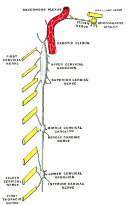
Diagram of the cervical sympathetic. (Labeled as "Upper cervical ganglion")
|
|
| Details | |
| Identifiers | |
| Latin | ganglion cervicale superius |
| MeSH | A08.340.315.350.850 |
| Dorlands /Elsevier |
g_02/12384383 |
| TA | A14.3.01.009 |
| FMA | 6467 |
|
Anatomical terms of neuroanatomy
[]
|
|
The superior cervical ganglion (SCG) is part of the autonomic nervous system (ANS) responsible for maintaining homeostasis of the body. More specifically it is part of the sympathetic nervous system, a division of the ANS most commonly associated with the fight or flight response. The ANS is composed of pathways that lead to and from ganglia, groups of nerve cells. A ganglion allows a large amount of divergence in a neuronal pathway and also enables a more localized circuitry for control of the innervated targets. The SCG is the only ganglion in the sympathetic nervous system that innervates the head and neck. It is the largest and most rostral (superior) of the three cervical ganglia. The SCG innervates many organs, glands and parts of the carotid system in the head.
The SCG is located opposite the second and third cervical vertebræ. It lies deep to the sheath of the internal carotid artery and internal jugular vein, and anterior to the Longus capitis muscle. The SCG contains neurons that supply sympathetic innervation to a number of target organs within the head.
The SCG also contributes to the cervical plexus. The cervical plexus is formed from a unification of the anterior divisions of the upper four cervical nerves. Each receives a gray ramus communicans from the superior cervical ganglion of the sympathetic trunk.
The superior cervical ganglion is a reddish-gray color, and usually shaped like a football with tapering ends. Sometimes the SCG is broad and flattened, and occasionally constricted at intervals. It formed by the coalescence of four ganglia, corresponding to the upper four cervical nerves, C1-C4. The bodies of these preganglionic sympathetic neurons are specifically located in the lateral horn of the spinal cord. These preganglionic neurons then enter the SCG and synapse with the postganglionic neurons that leave the rostral end of the SCG and innervate target organs of the head.
...
Wikipedia
