Stria terminalis
| Terminal stria | |
|---|---|

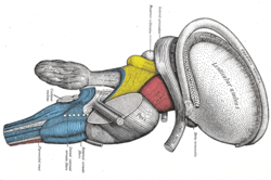
Dissection of brain-stem. Lateral view. (Stria terminalis labeled at upper right.)
|
|

Bed nucleus of the stria terminalis of the mouse brain
|
|
| Details | |
| Identifiers | |
| Latin | stria terminalis |
| NeuroNames | hier-268 |
| NeuroLex ID | Stria terminalis |
| TA | A14.1.09.275 |
| FMA | 61974 |
|
Anatomical terms of neuroanatomy
[]
|
|
The stria terminalis (or terminal stria) is a structure in the brain consisting of a band of fibers running along the lateral margin of the ventricular surface of the thalamus. Serving as a major output pathway of the amygdala, the stria terminalis runs from its centromedial division to the ventral medial nucleus of the hypothalamus.
The stria terminalis covers the thalamostriate vein, marking a line of separation between the thalamus and the caudate nucleus as seen upon gross dissection of the ventricles of the brain, viewed from the superior aspect.
The stria terminalis extends from the region of the interventricular foramina to the temporal horn of the lateral ventricle, carrying fibers from the amygdala to the septal nuclei, hypothalamic, and thalamic areas of the brain. It also carries fibers projecting from these areas back to the amygdala.
The activity of the bed nucleus of the stria terminalis correlates with anxiety in response to threat monitoring. It is thought to act as a relay site within the hypothalamic-pituitary-adrenal axis and regulate its activity in response to acute stress. However, the stress response is time related and the BNST does not activate for contextual fear. This means that a sudden scary situation that is under ten minutes long, does not activate the BNST. stress. It is also thought to promote behavioral inhibition in response to unfamiliar individuals, by input from the orbitofrontal cortex. Bilateral disruption of this pathway has been shown to attenuate reinstatement of drug seeking behaviour in rodents.
...
Wikipedia
