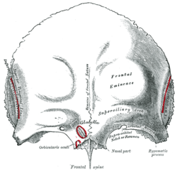
Squamous part of the frontal bone
| Squamous part of the frontal bone | |
|---|---|

Frontal bone. Outer surface. (The squamous part is the upper two thirds.)
|
|

Frontal bone. Inner surface. (The squamous part is the upper two thirds.)
|
|
| Details | |
| Identifiers | |
| Latin | Squama frontalis |
| TA | A02.1.03.002 |
| FMA | 52848 |
|
Anatomical terms of bone
[]
|
|
There are two surfaces of the squamous part of the frontal bone: the external surface, and the internal surface.
The external surface is convex and usually exhibits, in the lower part of the middle line, the remains of the frontal suture; in infancy this suture divides the frontal bone into two and later fuses. A condition where fusion has not taken place, may persist throughout life and is referred to as a metopic suture.
On either side of this suture, about 3 cm. above the supraorbital margin, is a rounded elevation, the frontal eminence (tuber frontale).
These eminences vary in size in different individuals, are occasionally unsymmetrical, and are especially prominent in young skulls; the surface of the bone above them is smooth, and covered by the galea aponeurotica.
Below the frontal eminences, and separated from them by a shallow groove, are two arched elevations, the superciliary arches; these are prominent medially, and are joined to one another by a smooth elevation named the glabella. They are larger in the male than in the female, and their degree of prominence depends to some extent on the size of the frontal air sinuses; prominent ridges are, however, occasionally associated with small air sinuses.
Beneath each superciliary arch is a curved and prominent margin, the supraorbital margin, which forms the upper boundary of the base of the orbit, and separates the squamous part from the orbital portion of the bone.
The lateral part of this margin is sharp and prominent, affording to the eye, in that situation, considerable protection from injury; the medial part is rounded.
At the junction of its medial and intermediate thirds is a notch, sometimes converted into a foramen, the supraorbital notch or foramen, which transmits the supraorbital vessels and nerve.
A small aperture in the upper part of the notch transmits a vein from the diploë to join the supraorbital vein.
...
Wikipedia
