Robustoxin
| Delta Atracotoxin | |||||||||
|---|---|---|---|---|---|---|---|---|---|

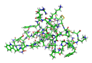
3D stick model of delta-atracotoxin-Ar1 (robustoxin)
|
|||||||||
| Identifiers | |||||||||
| Symbol | Atracotoxin | ||||||||
| Pfam | PF05353 | ||||||||
| InterPro | IPR008017 | ||||||||
| SCOP | 1qdp | ||||||||
| SUPERFAMILY | 1qdp | ||||||||
| OPM protein | 1vtx | ||||||||
|
|||||||||
| Available protein structures: | |
|---|---|
| Pfam | structures |
| PDB | RCSB PDB; PDBe; PDBj |
| PDBsum | structure summary |
Delta atracotoxin (δ-ACTX-Ar1, robustoxin, or robustotoxin) is a low-molecular-weight neurotoxic polypeptide found in the venom of the Sydney funnel-web spider (Atrax robustus).
Delta atracotoxin produces potentially fatal neurotoxic symptoms in primates by slowing the inactivation of sodium ion channels in autonomic and motor neurons. In the spiders' intended insect prey, the toxin exerts this same activity upon potassium and calcium ion channels.
The structure of atracotoxin comprises a core beta region with a cystine knot motif, a feature seen in other neurotoxic polypeptides.
Since 1927 records are kept of envenomations by the Sydney funnel-web spider and 14 deaths have been reported in medical literature between 1927 and 1981, when the antivenom became available. In all cases in which the sex of the spider was determined, death occurred after a bite from a male spider.
Delta atracotoxin is a 42-residue peptide toxin. The amino acid sequence of delta atracotoxin is unusual in that it contains three consecutive cysteine residues at positions 14–16. The amino acid sequence of delta atracotoxin is:
Cysteine bridges exist between Cys1 and Cys15, Cys8 and Cys20, Cys14 and Cys31, and Cys16 and Cys42.
The structure consists of a small triple-stranded beta-sheet stabilized by a disulfide knot, followed by a C-terminal extension comprising three classic or inverse y-turns. The disulfide knot is a ring consisting of two disulfide bonds (1-15 and 8-20) and the connecting backbone, through which a third disulfide bond (14-31) passes. The β-sheet, defined on the basis of inter-sheet hydrogen bonds, consists of residues 6-8 (strand I), 19-21 (strand II) and 29-32 (strand III), with a topology of +2x, —1. The two hydrogen bonds (one amide of which has a slowly exchanging amide proton) between strands I and III are distorted (NH to CO distance between 2.5 and 3.0 A). There are four hydrogen bonds between strands II and III (all of which have corresponding slowly exchanging amide protons), three being present in most of the structures and one in half of the structures. The structure contains a number of chain reversals. The first is not well defined and is either a type II ß-turn (Lys3-Asn6) or a y-turn centered on Arg5. Chain reversal II is a y turn centered on Gly9. Chain reversal III is not well defined, being either a type I ß-turn (Asnn-Cys14) or an inverse y-turn centered on Asn11. Chain reversal IV (Cys15-Met18) is not stabilized by a hydrogen bond but has a cis peptide bond between Cys16 and Pro17 and resembles a type Via turn. The fifth chain reversal occurs in the region of residues 22-28, which fulfill the criteria for an i2-loop. The C-terminal extension, stabilized by the Cys16-Cys42 disulfide bond, consists of three y-turns, VI-VIII, that are, respectively, an inverse turn, centered on Thr33, a classic turn centered on Ile35 and an inverse turn centered on Phe39. All three of the y-turn hydrogen bonds have slowly exchanging amide protons (although this is not the case for the other turns). The only slowly exchanging amide proton not accounted for by consensus hydrogen bonds in any secondary structure element is that of Gly37 (which hydrogen bonds to Thr34 in one of the structures). The conformations of the Cys1-Cys15 and Cys8-Cys20 disulfide bonds are well defined and have negative and positive Xss, respectively; the other two bonds have lower order parameters. The hydrophobic core of RBX is limited, consisting of essentially the disulfide knot cystine residues and the buried Met18. However, the 22-28 loop contains one apolar residue, Ala23, and three aromatics, Tyr22, Trp24 and Tyr25, and is flanked by Ile21 at its N-terminus and Trp7 near its C-terminus, so this region represents a significant non-polar surface on the molecule. RBX is highly positively charged, with one Arg (sequence position 5) and six Lys (3, 4, 10, 19, 40 and 41) residues, balanced only by Glu12 and Asp13. These charged residues form three patches on the surface. Patch A consists of the positively charged residues 3,4 and 5, patch B of residues 10, 12, 13 and the N-terminus (including possible salt bridges between Lys10 and Glu12 and Asp13 and the N-terminus), and patch C of 19, 40, 41 and the C-terminus.
...
Wikipedia
