Purkinje fibre
| Purkinje fibers | |
|---|---|

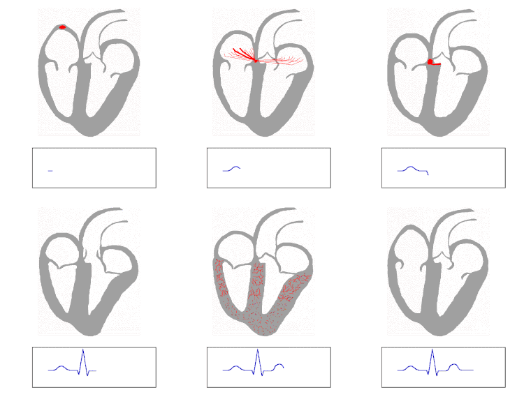
Isolated heart conduction system showing Purkinje fibers
|
|

The QRS complex is the large peak.
|
|
| Details | |
| Identifiers | |
| Latin | Rami subendocardiales |
| Dorlands /Elsevier |
f_05/12361434 |
| TA | A12.1.06.008 |
| FMA | 76538 |
|
Anatomical terminology
[]
|
|
The Purkinje fibers (/pərˈkɪndʒiː/ pər-KIN-jee) (Purkinje tissue or subendocardial branches) are located in the inner ventricular walls of the heart, just beneath the endocardium in a space called the subendocardium. The Purkinje fibers are specialised conducting fibers larger than cardiomyocytes with fewer myofibrils and a large number of that are able to conduct cardiac action potentials more quickly and efficiently than any other cells in the heart. Purkinje fibers allow the heart's conduction system to create synchronized contractions of its ventricles, and are, therefore, essential for maintaining a consistent heart rhythm.
Purkinje fibers are a unique cardiac end-organ. Further histologic examination reveals that these fibers are split in ventricles walls. The electrical origin of atrial Purkinje fibers arrives from the sinoatrial node.
Given no aberrant channels, the Purkinje fibers are distinctly shielded from each other by collagen or the cardiac skeleton.
The Purkinje fibers are further specialized to rapidly conduct impulses (numerous fast voltage-gated sodium channels and , fewer myofibrils than the surrounding muscle tissue). Purkinje fibers take up stain differently from the surrounding muscle cells because of relatively fewer myofibrils than other cardiac cells and the presence of glycogen around the nucleus causes Purkinje fibers to appear, on a slide, lighter and larger than their neighbors, arranged along the longitudinal direction (parallel to the cardiac vector) . They are often binucleated cells.
...
Wikipedia
