Presenilin
| Presenilin | |||||||||
|---|---|---|---|---|---|---|---|---|---|

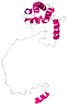
Solution structure of the human presenilin-1 CTF subunit.
|
|||||||||
| Identifiers | |||||||||
| Symbol | Presenilin | ||||||||
| Pfam | PF01080 | ||||||||
| Pfam clan | CL0130 | ||||||||
| InterPro | IPR001108 | ||||||||
| MEROPS | A22 | ||||||||
| TCDB | 1.A.54 | ||||||||
| OPM superfamily | 277 | ||||||||
| OPM protein | 4hyg | ||||||||
|
|||||||||
| Available protein structures: | |
|---|---|
| Pfam | structures |
| PDB | RCSB PDB; PDBe; PDBj |
| PDBsum | structure summary |
| presenilin 1 (Alzheimer's disease 3) |
|
|---|---|
| Identifiers | |
| Symbol | PSEN1 |
| Alt. symbols | AD3 |
| Entrez | 5663 |
| HUGO | 9508 |
| OMIM | 104311 |
| RefSeq | NM_000021 |
| UniProt | P49768 |
| Other data | |
| EC number | 3.4.23.- |
| Locus | Chr. 14 q24.3 |
| presenilin 2 (Alzheimer's disease 4) |
|
|---|---|
| Identifiers | |
| Symbol | PSEN2 |
| Alt. symbols | AD4 |
| Entrez | 5664 |
| HUGO | 9509 |
| OMIM | 600759 |
| RefSeq | NM_000447 |
| UniProt | P49810 |
| Other data | |
| EC number | 3.4.23.- |
| Locus | Chr. 1 q31-q42 |
Presenilins are a family of related multi-pass transmembrane proteins which constitute the catalytic subunits of the gamma-secretase intramembrane protease complex. They were first identified in screens for mutations causing early onset forms of familial Alzheimer's Disease by Peter St George-Hyslop at the Centre for Research in Neurodegenerative Diseases at the University of Toronto, and now also at the University of Cambridge. Vertebrates have two presenilin genes, called PSEN1 (located on chromosome 14 in humans) that encodes presenilin 1 (PS-1) and PSEN2 (on chromosome 1 in humans) that codes for presenilin 2 (PS-2). Both genes show conservation between species, with little difference between rat and human presenilins. The nematode worm C. elegans has two genes that resemble the presenilins and appear to be functionally similar, sel-12 and hop-1.
Presenilins undergo cleavage in an alpha helical region of one of the cytoplasmic loops to produce a large N-terminal and a smaller C-terminal fragment that together form part of the functional protein. Cleavage of presenilin 1 can be prevented by a mutation that causes the loss of exon 9, and results in loss of function. Presenilins play a key role in the modulation of intracellular Ca2+ involved in presynaptic neurotransmitter release and long-term potentiation induction.
The structure of presenilin-1 is still controversial, although recent research has produced a more widely accepted model. When first discovered, the PSEN1 gene was subjected to hydrophobicity analysis that predicted that the protein would contain ten trans-membrane domains. All previous models agreed that the first six putative membrane-spanning regions cross the membrane. These regions correspond to the N-terminal fragment of PS-1 but the structure of the C-terminal fragment was disputed. A recent paper by Spasic et al. provides strong evidence of a nine transmembrane structure with cleavage and assembly into the gamma-secretase complex prior to insertion into the plasma membrane. However, because this is a protein with large numbers of hydrophobic regions, it is unlikely that x-ray crystallography will provide definitive proof of the structure.
...
Wikipedia
