Posterior parietal cortex
| Posterior parietal cortex | |
|---|---|

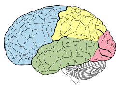
Lobes of the brain. Parietal lobe is yellow, and the posterior portion is near the red region.
|
|

Lateral surface of the brain with Brodmann's areas numbered. (#5 and #7 in upper right)
|
|
| Details | |
| Latin | Cortex parietalis posterior |
|
Anatomical terms of neuroanatomy
[]
|
|
The posterior parietal cortex (the portion of parietal neocortex posterior to the primary somatosensory cortex) plays an important role in planned movements, spatial reasoning, and attention.
Damage to the posterior parietal cortex can produce a variety of sensorimotor deficits, including deficits in the perception and memory of spatial relationships, inaccurate reaching and grasping, in the control of eye movement, and inattention. The two most striking consequences of PPC damage are apraxia and hemispatial neglect.
The posterior parietal cortex receives input from the three sensory systems that play roles in the localization of the body and external objects in space: the visual system, the auditory system, and the somatosensory system. In turn, much of the output of the posterior parietal cortex goes to areas of frontal motor cortex: the dorsolateral prefrontal cortex, various areas of the secondary motor cortex, and the frontal eye field.
The posterior parietal cortex is divided by the intraparietal sulcus to form the dorsal superior parietal lobule and the ventral inferior parietal lobule. Brodmann area 7 is part of the superior parietal lobule, but some sources include Brodmann area 5. The inferior parietal lobule is further subdivided into the supramarginal gyrus, the temporoparietal junction, and the angular gyrus. The inferior parietal lobule corresponds to Brodmann areas 39 and 40.
The posterior parietal cortex has been understood to have separate representations for different motor effectors (e.g. arm vs. eye).
In addition to separation based on effector type, some regions are activated during both decision and execution, while other regions are only active during execution. In one study, single cell recordings showed activity in parietal reach region while non-human primates decided whether to reach or make a saccade to a target, and activity persisted during the chosen movement if and only if the monkey chose to make a reaching movement. However, cells in area 5d were only active after the decision was made to reach with the arm. Another study found that neurons in area 5d only encoded the next movement in a sequence of reach movements, and not reach movements later in the sequence.
...
Wikipedia
