Pleural empyema
| Pleural empyema | |
|---|---|
| Synonyms | Pyothorax, purulent pleuritis |
 |
|
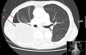
| CT chest showing large right sided hydro-pneumothorax from pleural empyema. Arrows A: air, B: fluid | |
| Classification and external resources | |
| Specialty | pulmonology |
| ICD-10 | J86 |
| ICD-9-CM | 510 |
| DiseasesDB | 4200 |
| MedlinePlus | 000123 |
| eMedicine | med/659 |
| MeSH | D016724 |
Pleural empyema is empyema (an accumulation of pus) in the pleural cavity that can develop when bacteria invade the pleural space, usually in the context of a pneumonia. It is one of various kinds of pleural effusion. There are three stages: exudative, when there is an increase in pleural fluid with or without the presence of pus; fibrinopurulent, when fibrous septa form localized pus pockets; and the final organizing stage, when there is scarring of the pleura membranes with possible inability of the lung to expand. Simple pleural effusions occur in up to 40% of bacterial pneumonias. They are usually small and resolve with appropriate antibiotic therapy. If however an empyema develops additional intervention is required.
The clinical presentation of both the adult and pediatric patient with pleural empyema depends upon several factors, including the causative micro-organism. Most cases present themselves in the setting of a pneumonia, although up to one third of patients do not have clinical signs of pneumonia and as many as 25% of cases are associated with trauma (including surgery). Typical symptoms include cough, chest pain, shortness of breath and fever.
The initial investigations for suspected empyema remains chest X-ray, although it cannot differentiate an empyema from uninfected parapneumonic effusion.Ultrasound must be used to confirm the presence of a pleural fluid collection and can be used to estimate the size of the effusion, differentiate between free and loculated pleural fluid and guide thoracocentesis if necessary. Chest CT and MRI do not provide additional information in most cases and should therefore not be performed routinely. On a CT scan, empyema fluid most often has a radiodensity of about 0-20 Hounsfield units (HU), but gets over 30 HU when becoming more thickened with time.
...
Wikipedia
