Patellar tendon rupture
| Patellar tendon rupture | |
|---|---|
 |
|
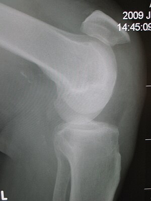
| Patellar tendon rupture showing a marked distance between the tibial tuberosity and the bottom of the patella. | |
| Classification and external resources | |
| Specialty | emergency medicine |
| ICD-10 | S76.1 |
| ICD-9-CM | 727.66 |
| eMedicine | article/1249472 |
Patellar tendon rupture is a rupture of the tendon that connects the patella to the tibia. The superior portion of the patellar tendon attaches on the posterior portion of the patella, and the posterior portion of the patella tendon attaches to the tibial tubercle on the front of the tibia. Above the patella are the quadriceps muscle (large muscles on the front of the thigh), the quadriceps tendon attaches to the top of the patella. This structure allows the knee to flex and extend, allowing use of basic functions such as walking and running.
There are three possible forms of patellar tendon rupture. The first form of rupture is a complete tear. In a complete tear, the tendon separates completely from the top of the tibia which results in the inability to straighten one's leg. When the tendon tears, it can break a piece of the bone off of the kneecap. The second form of rupturing the patellar tendon is a partial tear. A partial tear is when some of the fibers of the patellar tendon are torn but the majority of the tendon is still attached to the soft tissue located at the posterior end of the patella bone. The third form of rupture is caused by patellar tendinitis ("jumper's knee"). Patellar tendinitis causes the tendon to be torn in the middle due to the tissue damage it has been acquiring from over-use. Patellar tendonitis is inflammation of the tendon which results in the weakening of the tendon. Tendonitis is caused by excessive jumping or running without sufficient rest.
The tell-tale sign of a ruptured patella tendon is the movement of the patella further up the quadriceps. When rupture occurs, the patella loses support from the tibia and moves toward the hip when the quadriceps muscle contracts, hindering the leg's ability to extend. This means that those affected cannot stand, as their knee buckles and gives way when they attempt to do so.
Patellar tendon rupture can usually be diagnosed by physical examination. The most common signs are: tenderness, the tendon's loss of tone, loss of ability to raise the straight leg and observation of the high-riding patella. Radiographically, patella alta can be detected using the Insall and Salvati method when the patella is shorter than its tendon. Partial tears may be visualized using MRI scans.
Patellar tendon rupture must be treated surgically. With a tourniquet applied, the tendon was exposed through a midline longitudinal incision extending from the upper patellar pole to the tibial tuberosity. The tendon either avulsed (detached) from the lower patellar pole or lacerated. Even so, continuity and tone of the tendon should be restored taking in consideration the patellar height.
...
Wikipedia
