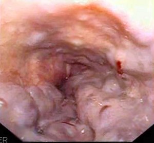
Oesophageal varices
| Esophageal varices | |
|---|---|
 |
|
| Gastroscopy image of esophageal varices with prominent cherry-red spots | |
| Classification and external resources | |
| Specialty | Gastroenterology |
| ICD-10 | I85 |
| ICD-9-CM | 456.0-456.2 |
| DiseasesDB | 9177 |
| MedlinePlus | 000268 |
| eMedicine | med/745 radio/269 |
| MeSH | D004932 |
Esophageal varices (sometimes spelled oesophageal varices) are extremely dilated sub-mucosal veins in the lower third of the esophagus. They are most often a consequence of portal hypertension, commonly due to cirrhosis; patients with esophageal varices have a strong tendency to develop bleeding. Esophageal varices are typically diagnosed through an esophagogastroduodenoscopy.
The upper two thirds of the esophagus is drained via the esophageal veins, which carry deoxygenated blood from the esophagus to the azygos vein, which in turn drains directly into the superior vena cava. These veins have no part in the development of esophageal varices. The lower one third of the esophagus is drained into the superficial veins lining the esophageal mucosa, which drain into the left gastric vein (coronary vein), which in turn drains directly into the portal vein. These superficial veins (normally only approximately 1 mm in diameter) become distended up to 1–2 cm in diameter in association with portal hypertension.
Normal portal pressure is approximately 9 mmHg compared to an inferior vena cava pressure of 2–6 mmHg. This creates a normal pressure gradient of 3–7 mmHg. If the portal pressure rises above 12 mmHg, this gradient rises to 7–10 mmHg. A gradient greater than 5 mmHg is considered portal hypertension. At gradients greater than 10 mmHg, blood flow through the hepatic portal system is redirected from the liver into areas with lower venous pressures. This means that collateral circulation develops in the lower esophagus, abdominal wall, stomach, and rectum. The small blood vessels in these areas become distended, becoming more thin-walled, and appear as varicosities.
...
Wikipedia
