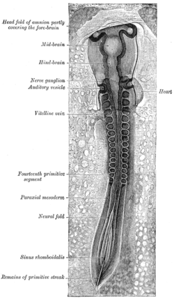
Neural folds
| Neural fold | |
|---|---|

Chick embryo of thirty-three hours’ incubation, viewed from the dorsal aspect. 30x. (Neural fold labeled at center left, third from the bottom.)
|
|
| Details | |
| Carnegie stage | 9 |
| Precursor | neural plate |
| Gives rise to | neural tube |
| Identifiers | |
| Latin | plica neuralis |
| Code | TE E5.13.1.0.1.0.2 |
|
Anatomical terminology
[]
|
|
The neural fold is a structure that arises during neurulation in the embryonic development of both birds and mammals among other organisms. This structure is associated with primary neurulation, meaning that it forms by the coming together of tissue layers, rather than a clustering, and subsequent hollowing out, of individual cells (known as secondary neurulation). In humans, the neural folds are responsible for the formation of the anterior end of the neural tube. The neural folds are derived from the neural plate, a preliminary structure consisting of elongated ectoderm cells. The folds give rise to neural crest cells, as well as bringing about the formation of the neural tube.
In the embryo, the formation of the neural folds originates from the area where the neural plate and the surrounding ectoderm converge. This region of the embryo is formed after gastrulation, and consists of epithelial tissue. Here, the epithelial cells elongate by means of microtubule polymerization, increasing their height. The thumbnail below shows this process, as well as the subsequent formation of the neural crest cells and the neural tube, which arise from the joining of the neural folds.
The formation of the neural fold is initiated by the release of calcium from within the cells. The released calcium interacts with proteins that can modify the actin filaments in the outer epithelial tissue, or ectoderm, in order to induce the dynamic cell movements necessary to create the fold. These cells are held together by cadherins (specifically E and N-cadherin), types of intercellular binding protein. When the cells at the peaks of the neural folds come in proximity with each other, it is the affinity for similar cadherin molecules (N-cadherins) that allows these cells to bind to each other. Thus, when the neural tube precursor cells begin expressing N-cadherin in the place of E-cadherin, this causes the neural tube to form and separate from the ectoderm and settle inside the embryo. When the cells fail to associate in a manner that is not part of the normal course of development, severe diseases can occur.
...
Wikipedia
