Meniscus (anatomy)
| Meniscus | |
|---|---|

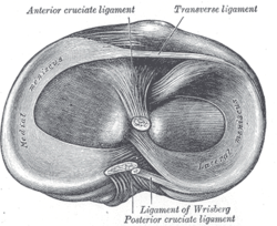
Head of right tibia seen from above, showing menisci and attachments of ligaments
|
|

Left knee-joint from behind, showing interior ligaments
|
|
| Details | |
| Identifiers | |
| Latin | Menisci |
| TA | A03.0.00.033 |
| FMA | 76690 |
|
Anatomical terminology
[]
|
|
In anatomy, a meniscus (from Greek meniskos, "crescent") is a crescent-shaped fibrocartilaginous structure that, in contrast to an articular disk, only partly divides a joint cavity. In humans they are present in the knee, wrist, acromioclavicular, sternoclavicular, and temporomandibular joints; in other animals they may be present in other joints.
Generally, the term 'meniscus' is used to refer to the cartilage of the knee, either to the lateral or medial meniscus. Both are cartilaginous tissues that provide structural integrity to the knee when it undergoes tension and torsion. The menisci are also known as "semi-lunar" cartilages — referring to their half-moon, crescent shape.
The menisci of the knee are two pads of fibrocartilaginous tissue which serve to disperse friction in the knee joint between the lower leg (tibia) and the thigh (femur). They are on the top and flat on the bottom, articulating with the tibia. They are attached to the small depressions (fossae) between the condyles of the tibia (intercondyloid fossa), and towards the center they are unattached and their shape narrows to a thin shelf. The blood flow of the meniscus is from the periphery (outside) to the central meniscus. Blood flow decreases with age and the central meniscus is avascular by adulthood, leading to very poor healing rates.
...
Wikipedia
