Malleolus
| malleolus | |
|---|---|

Coronal section through right talocrural and talocalcaneal joints.
|
|

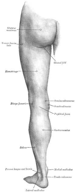
Back of left lower extremity. (Medial malleolus labeled at bottom right.)
|
|
| Details | |
| Identifiers | |
| Latin | malleolus, malleoli |
| TA |
A02.5.06.020 A02.5.07.014 |
|
Anatomical terms of bone
[]
|
|
A malleolus is the bony prominence on each side of the human ankle.
Each leg is supported by two bones, the tibia on the inner side (medial) of the leg and the fibula on the outer side (lateral) of the leg. The medial malleolus is the prominence on the inner side of the ankle, formed by the lower end of the tibia. The lateral malleolus is the prominence on the outer side of ankle, formed by the lower end of the fibula.
The medial surface of the lower extremity of tibia is prolonged downward to form a strong pyramidal process, flattened from without inward - the medial malleolus.
Structures that pass behind medial malleolus deep to flexor retinaculum:
The lower extremity of the fibula, also called the distal extremity or external malleolus, is of a pyramidal form and somewhat flattened from side to side; it descends to a lower level than the medial malleolus.
Bones of the right leg. Anterior surface.
Back of left lower extremity.
Lateral aspect of right leg.
Dorsum of Foot. Ankle joint. Deep dissection
Dorsum of Foot. Ankle joint. Deep dissection
Ankle joint. Deep dissection. Anterior view.
Ankle joint. Deep dissection. Lateral view.
Ankle joint. Deep dissection.
Ankle joint. Deep dissection.
Ankle joint. Deep dissection.
Ankle joint. Deep dissection.
Ankle joint. Deep dissection.
Ankle and tarsometarsal joints. Bones of foot.Deep dissection.
Ankle and tarsometarsal joints. Bones of foot.Deep dissection.
Ankle joint. Bones of foot.Deep dissection.
Ankle and tarsometatarsal joint. Deep dissection.Anterior view
This article incorporates text in the public domain from the 20th edition of Gray's Anatomy (1918)
...
Wikipedia
