
Magnetic resonance images
| Magnetic resonance imaging | |
|---|---|
| Medical diagnostics | |
| Synonyms | nuclear magnetic resonance imaging (NMRI), magnetic resonance tomography (MRT) |
| ICD-9-CM | 88.91 |
| MeSH | D008279 |
| MedlinePlus | 003335 |
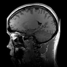
Clinical magnetic resonance imaging (MRI) is a technique to form pictures of the anatomy and the processes of living organisms. MRI scanners use strong magnetic fields, radio waves, and field gradients to generate images. MRI does not involve x-rays, which distinguishes it from computed tomography (CT or CAT). MRI is used extensively in medical imaging in radiology, to study the body in both health and disease.
While the hazards of x-rays are now well-controlled in most medical contexts, MRI still may be seen as superior to CT in this regard. MRI is widely used in hospitals and clinics for medical diagnosis, staging of disease and follow-up without exposing the body to ionizing radiation. MRI often may yield different diagnostic information compared with CT. There may be risks and discomfort associated with MRI scans. Compared with CT, MRI scans typically take greater time, are louder, and usually require that the subject go into a narrow, confining tube. In addition, people with some medical implants or other non-removable metal inside the body may be unable to undergo an MRI examination safely.
MRI was originally called 'NMRI' (nuclear magnetic resonance imaging). It is based upon the science of nuclear magnetic resonance (NMR). Certain atomic nuclei are able to absorb and emit radio frequency energy when placed in an external magnetic field. In clinical and research MRI, hydrogen atoms are most often used to generate a detectable radio-frequency signal that is received by antennas in close proximity to the anatomy being examined. Hydrogen atoms exist naturally in people and other biological organisms in abundance, particularly in water and fat. For this reason, most MRI scans essentially map the location of water and fat in the body. Pulses of radio waves excite the nuclear spin energy transition, and magnetic field gradients localize the signal in space. By varying the parameters of the pulse sequence, different contrasts may be generated between tissues based on the relaxation properties of the hydrogen atoms therein.
...
Wikipedia