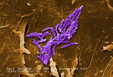
M. fortuitum
| Mycobacterium fortuitum | |
|---|---|
 |
|
| A scanning electron micrograph of Mycobacterium fortuitum | |
| Scientific classification | |
| Kingdom: | Bacteria |
| Phylum: | Actinobacteria |
| Order: | Actinomycetales |
| Suborder: | Corynebacterineae |
| Family: | Mycobacteriaceae |
| Genus: | Mycobacterium |
| Species: | M. fortuitum |
| Binomial name | |
|
Mycobacterium fortuitum Da Costa Cruz 1938, ATCC 6841 |
|
Mycobacterium fortuitum is a nontuberculous species of the phylum actinobacteria (Gram-positive bacteria with high guanine and cytosine content, one of the dominant phyla of all bacteria), belonging to the genus mycobacterium.
Mycobacterium fortuitum is a fast-growing species that can cause infections. The term "fast growing" is a reference to a growth rate of 3 or 4 days, when compared to other Mycobacteria that may take weeks to grow out on laboratory media. Pulmonary infections of M. fortuitum are uncommon, but Mycobacterium fortuitum can cause local skin disease, osteomyelitis (inflammation of the bone), joint infections and infections of the eye after trauma. Mycobacterium fortuitum has a worldwide distribution and can be found in natural and processed water, sewage, and dirt.
Bacteria classified as Mycobacteria, include the causative agents for tuberculosis and leprosy. Mycobacteria are sometimes referred to as “acid-fast bacteria,” a term referencing their response to a laboratory staining technique. This simply means that when microscopic slides of these bacteria are rinsed with an acidic solution, they retain a red dye. Mycobacterium fortuitum is one of the many species of nontuberculosis mycobacteria (NTM) that are commonly found in the environment. These are not involved in tuberculosis. This does not mean, however, that they will not cause an infection in the right circumstances.
M. fortuitum infection can be a nosocomial (hospital acquired) disease. Surgical sites may become infected after the wound is exposed directly or indirectly to contaminated tap water. Other possible sources of M. fortuitum infection include implanted devices such as catheters, injection site abscesses, and contaminated endoscopes. Recent publication on Rapidly Growing Mycobacteria (RGM) is available provides the following aspects of RGM: (i) its sources, predisposing factors, clinical manifestations, and concomitant fungal infections; (ii) the risks of misdiagnoses in the management of RGM infections in dermatological settings; (iii) the diagnoses and outcomes of treatment responses in common and uncommon infections in immunocompromised and immunocompetent patients; (iv) conventional versus current molecular methods for the detection of RGM; (v) the basic principles of a promising MALDI-TOF MS, sampling protocol for cutaneous or subcutaneous lesions and its potential for the precise differentiation of M. fortuitum, M. chelonae, and M. abscessus; and (vi) improvements in RGM infection management as described in the recent 2011 Clinical and Laboratory Standards Institute (CLSI) guidelines, including interpretation criteria of molecular methods and antimicrobial drug panels and their break points [minimum inhibitory concentrations (MICs)], which have been highlighted for the initiation of antimicrobial therapy (Kothavade RJ et al., 2012).
...
Wikipedia
