Limbic encephalopathy
| Limbic encephalitis | |
|---|---|
 |
|
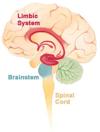
| The limbic system within the brain. | |
| Classification and external resources | |
| Specialty | neurology |
| ICD-10 | G04.81 |
| ICD-9-CM | 323.81 |
| DiseasesDB | 30707 |
| MeSH | D020363 |
Limbic encephalitis is a form of encephalitis, a disease characterised by inflammation of the brain. Limbic encephalitis is caused by autoimmunity: an abnormal state where the body produces antibodies against itself. Some cases are associated with cancer and some are not. Although the disease is known as "limbic" encephalitis, it is seldom limited to the limbic system and post-mortem studies usually show involvement of other parts of the brain. The disease was first described by Brierley and others in 1960 as a series of three cases. The link to cancer was first noted in 1968 and confirmed by later investigators.
The majority of cases of limbic encephalitis are associated with a tumour (diagnosed or undiagnosed). In cases caused by tumour, cure is only achieved when the tumour is removed completely (this is not always possible). Limbic encephalitis is classified according to the auto-antibody that causes the disease.
The most common types are:
Since 1999 following a report of case of a 15 -year-old teenager of Indian descent from South Africa, subacutely developed memory loss subsequent to herpes simplex type 1 encephalitis similar cases of non paraneoplastic LE its association with auto-antibody and response to steroid has been described. Limbic encephalitis associated with voltage‐gated potassium channel antibodies (VGKC‐Abs) may frequently be non‐paraneoplastic. A recent study of 15 cases of limbic encephalitis found raised VGKC‐Abs associated with non‐paraneoplastic disorders and remission following immunosuppressive treatment.
Symptoms develop over days or weeks. The subacute development of short-term memory deficits is considered the hallmark of this disease, but this symptom is often overlooked, because it is overshadowed by other more obvious symptoms such as headache, irritability, sleep disturbance, delusions, hallucinations, agitation, seizures and psychosis, or because the other symptoms mean the patient has to be sedated, and it is not possible to test memory in a sedated patient.
Examination of cerebrospinal fluid (CSF) shows elevated numbers of lymphocytes (but usually < 100 cells/µl); elevated CSF protein (but usually <1.5 g/l), normal glucose, elevated IgG index and oligoclonal bands. Patients with antibodies to voltage-gated potassium channels may have a completely normal CSF examination.
MRI brain is the mainstay of initial investigation pointing to limbic lobe pathology revealing increased T2 signal involving one or both temporal lobes in most cases.
...
Wikipedia
