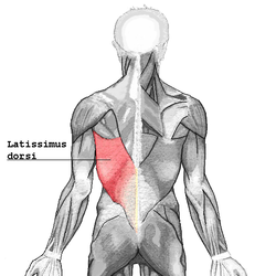
Latissimus dorsi muscle
| Latissimus Dorsi | |
|---|---|

Latissimus dorsi
|
|

Muscles connecting the upper extremity to the vertebral column.
|
|
| Details | |
| Origin | Spinous processes of vertebrae T7-T12, thoracolumbar fascia, iliac crest, inferior 3 or 4 ribs and inferior angle of scapula |
| Insertion | Floor of intertubercular groove of the humerus |
| Artery | Thoracodorsal branch of the subscapular artery |
| Nerve | Thoracodorsal nerve(C6,C7,C8) |
| Actions | Adducts, extends and internally rotates the arm when the insertion is moved towards the origin. When observing the muscle action of the origin towards the insertion, the lats are a very powerful rotator of the trunk. |
| Antagonist | Deltoid and trapezius muscle |
| Identifiers | |
| Latin | Musculus latissimus dorsi |
| Dorlands /Elsevier |
m_22/12549548 |
| TA | A04.3.01.006 |
| FMA | 13357 |
|
Anatomical terms of muscle
[]
|
|
The latissimus dorsi (/ˌləˈtɪsᵻməs ˈdɔːrsaɪ/) (plural: latissimi dorsi), meaning 'broadest [muscle] of the back' (Latin latus meaning 'broad', latissimus meaning 'broadest' and dorsum meaning the back), is the larger, flat, dorso-lateral muscle on the trunk, posterior to the arm, and partly covered by the trapezius on its median dorsal region. Latissimi dorsi are commonly known as "lats", especially among bodybuilders.
The latissimus dorsi is responsible for extension, adduction, transverse extension also known as horizontal abduction, flexion from an extended position, and (medial) internal rotation of the shoulder joint. It also has a synergistic role in extension and lateral flexion of the lumbar spine.
Due to bypassing the scapulothoracic joints and attaching directly to the spine, the actions the latissimi dorsi have on moving the arms can also influence the movement of the scapulae, such as their downward rotation during a pull up.
The number of dorsal vertebrae to which it is attached varies from four to eight; the number of costal attachments varies; muscle fibers may or may not reach the crest of the ilium.
A muscular slip, the axillary arch, varying from 7 to 10 cm in length, and from 5 to 15 mm in breadth, occasionally springs from the upper edge of the latissimus dorsi about the middle of the posterior fold of the axilla, and crosses the axilla in front of the axillary vessels and nerves, to join the under surface of the tendon of the pectoralis major, the coracobrachialis, or the fascia over the biceps brachii. This axillary arch crosses the axillary artery, just above the spot usually selected for the application of a ligature, and may mislead a surgeon. It is present in about 7% of the population and may be easily recognized by the transverse direction of its fibers. Guy et al. extensively described this muscular variant using MRI data and positively correlated its presence with symptoms of neurological impingement.
...
Wikipedia
