Ischial tuberosities
| Ischial tuberosity | |
|---|---|

Capsule of hip-joint (distended). Posterior aspect. (Ischial tuberosity visible at bottom left.)
|
|

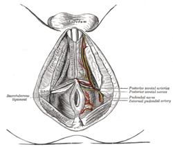
The superficial branches of the internal pudendal artery. (Ischial tuberosity visible at center left.)
|
|
| Details | |
| Identifiers | |
| Latin | Tuber ischiadicum, tuberositas ischiadica |
| Dorlands /Elsevier |
t_21/12827506 |
| TA | A02.5.01.204 |
| FMA | 17010 |
|
Anatomical terms of bone
[]
|
|
The ischial tuberosity (or tuberosity of the ischium, tuber ischiadicum), also known informally as the sit bones, or as a pair the sitting bones is a large swelling posteriorly on the superior ramus of the ischium. It marks the lateral boundary of the pelvic outlet.
When sitting, the weight is frequently placed upon the ischial tuberosity. The gluteus maximus provides cover in the upright posture, but leaves it free in the seated position.
The tuberosity is divided into two portions: a lower, rough, somewhat triangular part, and an upper, smooth, quadrilateral portion.
Muscles of the gluteal and posterior femoral regions, with ischial tuberosity highlighted in red.
Right hip bone. External surface.
Right hip bone. Internal surface.
Plan of ossification of the hip bone.
Diameters of inferior aperture of lesser pelvis (female).
Right hip-joint from the front.
The Obturator externus.
Anterior view of the pelvis with the ischial tuberosity labelled in the lower part of the image
This article incorporates text in the public domain from the 20th edition of Gray's Anatomy (1918)
...
Wikipedia
