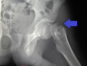
Intertrochanteric
| Hip fracture | |
|---|---|
| Synonyms | Proximal femur fracture |
 |
|
| Intertrochanteric hip fracture in a 17-year-old male | |
| Symptoms | Pain around the hip particularly with movement, shortening of the leg |
| Types | Intracapsular, extracapsular (intertrochanteric, subtrochanteric, greater trochanteric, lesser trochanteric) |
| Causes | Trauma such as a fall |
| Risk factors | Osteoporosis, taking many medications, alcohol use, metastatic cancer |
| Diagnostic method | X-ray, MRI, CT scan, bone scan |
| Differential diagnosis | Osteoarthritis, avascular necrosis of the hip, hernia, trochanteric bursitis |
| Prevention | Improved lighting, removal of loose rugs, exercise, treatment of osteoporosis |
| Treatment | Surgery |
| Medication | Opioids, nerve block |
| Prognosis | ~20% one year risk of death (older people) |
| Frequency | ~15% of women at some point |
| Classification | |
|---|---|
| External resources |
|
A hip fracture is a break that occurs in the upper part of the femur (thigh bone). Symptoms may include pain around the hip particularly with movement and shortening of the leg. Usually the person cannot walk.
They most often occur as a result of a fall. Risk factors include osteoporosis, taking many medications, alcohol use, and metastatic cancer. Diagnosis is generally by X-rays.Magnetic resonance imaging, a CT scan, or a bone scan may occasionally be required to make the diagnosis.
Pain management may occur with opioids or a nerve block. If a person's health is sufficient, surgery is generally recommended within two days. Options for surgery may include a total hip replacement or screws. Efforts to prevent deep vein thrombosis following surgery are recommended.
About 15% of women break their hip at some point in their life. Women are more often affected than men. Hip fractures become more common with age. The risk of death in the year following a fracture is about 20% in older people.
The classic clinical presentation of a hip fracture is an elderly patient who sustained a low-energy fall and now has groin pain and is unable to bear weight. Pain may be referred to the supracondylar knee. On examination, the affected extremity is often shortened and unnaturally, externally rotated compared to the unaffected leg.
Hip fracture following a fall is likely to be a pathological fracture. The most common causes of weakness in bone are:
The hip joint, an enarthrodial joint, can be described as a ball and socket joint. The femur connects at the acetabulum of the pelvis and projects laterally before angling medially and inferiorly to form the knee. Although this joint has three degrees of freedom, it is still stable due to the interaction of ligaments and cartilage. The labrum lines the circumference of the acetabulum to provide stability and shock absorption. Articular cartilage covers the concave area of acetabulum, providing more stability and shock absorption. Surrounding the entire joint itself is a capsule secured by the tendon of the psoas muscle and three ligaments. The iliofemoral, or Y, ligament is located anteriorly and serves to prevent hip hyperextension. The pubofemoral ligament is located anteriorly just underneath the iliofemoral ligament and serves primarily to resist abduction, extension, and some external rotation. Finally the ischiofemoral ligament on the posterior side of the capsule resists extension, adduction, and internal rotation. When considering the biomechanics of hip fractures, it is important to examine the mechanical loads the hip experiences during low energy falls.
...
Wikipedia
