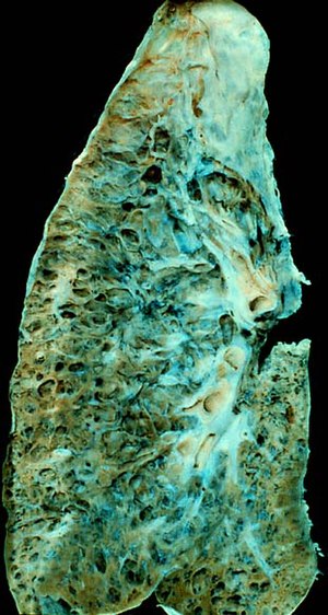
Interstitial pneumonitis
| Interstitial lung disease | |
|---|---|
 |
|
| End-stage pulmonary fibrosis of unknown origin, taken from an autopsy in the 1980s | |
| Classification and external resources | |
| Specialty | Pulmonology |
| ICD-10 | J84.9 |
| ICD-9-CM | 518.89, 508.1, 515, 516.3, 714.81, 770.7 |
Interstitial lung disease (ILD), or diffuse parenchymal lung disease (DPLD), is a group of lung diseases affecting the interstitium (the tissue and space around the air sacs of the lungs). It concerns alveolar epithelium, pulmonary capillary endothelium, basement membrane, perivascular and perilymphatic tissues. It may occur when an injury to the lungs triggers an abnormal healing response. Ordinarily, the body generates just the right amount of tissue to repair damage. But in interstitial lung disease, the repair process goes awry and the tissue around the air sacs (alveoli) becomes scarred and thickened. This makes it more difficult for oxygen to pass into the bloodstream. The term ILD is used to distinguish these diseases from obstructive airways diseases.
In children, several unique forms of ILD exist which are specific for the young age groups. The acronym chILD is used for this group of diseases and is derived from the English name, Children’s Interstitial Lung Diseases – chILD.
Prolonged ILD may result in pulmonary fibrosis, but this is not always the case. Idiopathic pulmonary fibrosis is interstitial lung disease for which no obvious cause can be identified (idiopathic), and is associated with typical findings both radiographic (basal and pleural based fibrosis with honeycombing) and pathologic (temporally and spatially heterogeneous fibrosis, histopathologic honeycombing and fibroblastic foci).
In 2013 interstitial lung disease affected 595,000 people globally. This resulted in 471,000 deaths.
ILD may be classified according to the cause. One method of classification is as follows:
Investigation is tailored towards the symptoms and signs. A proper and detailed history looking for the occupational exposures, and for signs of conditions listed above is the first and probably the most important part of the workup in patients with interstitial lung disease. Pulmonary function tests usually show a restrictive defect with decreased diffusion capacity (DLCO).
...
Wikipedia
