Infraorbital portion
| Infraorbital nerve | |
|---|---|

Left orbicularis oculi, seen from behind. (Infraorbital nerve labeled at lower left.)
|
|

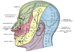
Sensory areas of the head, showing the general distribution of the three divisions of the fifth nerve. (Infraorbital nerve labeled at center left, at the nose.)
|
|
| Details | |
| From | maxillary nerve |
| Identifiers | |
| Latin | Nervus infraorbitalis |
| Dorlands /Elsevier |
n_05/12565913 |
| TA | A14.2.01.059 |
| FMA | 52978 |
|
Anatomical terms of neuroanatomy
[]
|
|
After the maxillary nerve enters the infraorbital canal, the nerve is frequently called the infraorbital nerve. This nerve innervates (sensory) the lower eyelid, upper lip, and part of the nasal vestibule and exits the infraorbital foramen of the maxilla. There is a cross innervation of this nerve on the other side of jaw.
The infraorbital nerve block is a type of local anesthetic nerve block used to induce analgesia in the distribution of the nerve for whatever purpose.
After a fracture of the floor of the orbit, the infraorbital nerve may become trapped, producing an area of anaesthesia under the orbital rim.
Mandibular division of the trigeminal nerve.
The nerves of the scalp, face, and side of neck.
Outline of side of face, showing chief surface markings.
An illustration of the path of the maxillary nerve.
Infaorbital and buccal nerve.Superficial dissection.Lateral view.
This article incorporates text in the public domain from the 20th edition of Gray's Anatomy (1918)
...
Wikipedia
