Ileocolic junction
| Ileocecal valve | |
|---|---|

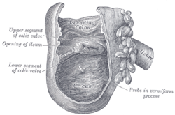
Interior of the cecum and lower end of ascending colon with the ileocecal valve labeled as "colic valve"
|
|

Endoscopic image of cecum with arrow pointing to ileocecal valve in foreground
|
|
| Details | |
| Artery | Ileocolic artery |
| Vein | Ileocolic vein |
| Identifiers | |
| Latin | Valva ileocaecalis or papilla ilealis |
| MeSH | D007080 |
| FMA | 15973 |
|
Anatomical terminology
[]
|
|
The ileocecal valve (ileal papilla, ileocaecal valve, Tulp's valve, Tulpius valve, Bauhin's valve, ileocecal eminence, valve of Varolius or colic valve) is a sphincter muscle valve that separates the small intestine and the large intestine. Its critical function is to limit the reflux of colonic contents into the ileum. Approximately two liters of fluid enters the colon daily through the ileocecal valve.
The ileocecal valve is distinctive because it is the only site in the gastrointestinal tract that is used for Vitamin B12 and bile acid absorption.
It was described by the Dutch physician Nicolaes Tulp (1593–1674), and thus it is sometimes known as Tulp's valve.
The valve was also described in 1588 by Gaspard Bauhin—hence the name Bauhin's Valve or Valve of Bauhin—in the preface of his first writing, De corporis humani partibus externis tractatus, hactenus non editus.
The histology of the ileocecal valve shows an abrupt change from a villous mucosa pattern of the ileum to a more colonic mucosa. A thickening of the muscularis mucosa, which is the smooth muscle tissue found beneath the mucosal layer of the digestive tract. A thickening of the muscularis externa is also noted.
There is also a variable amount of lymphatic tissue found at the valve.
The ileocecal valve has a papillose structure.
...
Wikipedia
