Hypothalamic–neurohypophyseal system
| Posterior pituitary | |
|---|---|

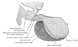
Pituitary gland. Posterior pituitary is in blue and Anterior pituitary is in orange. Pars nervosa and infundibular stalk are not labeled, but pars nervosa is at bottom and infundibular stalk is at top.
|
|

Median sagittal through the hypophysis of an adult monkey. (Posterior lobe labeled at bottom right.)
|
|
| Details | |
| Precursor | Neural tube (downward-growth of the diencephalon) |
| Artery | inferior hypophyseal artery |
| Vein | hypophyseal vein |
| Identifiers | |
| Latin | Pars nervosa glandulae pituitariae, pars nervosa hypophyseos, lobus posterior hypophyseos |
| MeSH | A06.407.747.734 |
| NeuroLex ID | Neurohypophosis |
| Dorlands /Elsevier |
Posterior pituitary hormones |
| TA | A11.1.00.006 |
| FMA | 74628 |
|
Anatomical terminology
[]
|
|
The posterior pituitary (or neurohypophysis) is the posterior lobe of the pituitary gland which is part of the endocrine system. The posterior pituitary is not glandular as is the anterior pituitary. Instead, it is largely a collection of axonal projections from the hypothalamus that terminate behind the anterior pituitary which serves as a site for the secretion of neurohypophysial hormones ( and vasopressin) directly into the blood. The hypothalamic–neurohypophyseal system is composed of the hypothalamus (the paraventricular nucleus and supraoptic nucleus), posterior pituitary, and these axonal projections.
The posterior pituitary consists mainly of neuronal projections (axons) of magnocellular neurosecretory cells extending from the supraoptic and paraventricular nuclei of the hypothalamus. These axons store and release neurohypophysial hormones and vasopressin into the neurohypohyseal capillaries, from there they get into the systemic circulation (and partly back into the hypophyseal portal system). In addition to axons, the posterior pituitary also contains pituicytes, specialized glial cells resembling astrocytes assisting in the storage and release of the hormones.
...
Wikipedia
