Hind-brain
| Hindbrain | |
|---|---|

|
|

Scheme of roof of fourth ventricle.
|
|
| Identifiers | |
| MeSH | A08.186.211.132.810 |
| NeuroNames | hier-531 |
| NeuroLex ID | Rhombencephalon |
| Dorlands /Elsevier |
r_12/12709581 |
| TA | A14.1.03.002 |
| FMA | 67687 |
|
Anatomical terms of neuroanatomy
[]
|
|
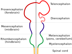
The hindbrain or rhombencephalon is a developmental categorization of portions of the central nervous system in vertebrates. It includes the medulla, pons, and cerebellum. Together they support vital bodily processes.
The hindbrain can be subdivided in a variable number of transversal swellings called rhombomeres. In the human embryo eight rhombomeres can be distinguished, from caudal to rostral: Rh8-Rh1. Rostrally, the isthmus demarcates the boundary with the midbrain.
A rare disease of the rhombencephalon—"rhombencephalosynapsis"—is characterized by a missing vermis resulting in a fused cerebellum. Patients generally present with cerebellar ataxia.
The caudal rhombencephalon has been generally considered as the initiation site for neural tube closure.
Rhombomeres Rh3-Rh1 form the metencephalon.
The metencephalon is composed of the pons and the cerebellum; it contains:
Rhombomeres Rh8-Rh4 form the myelencephalon.
The myelencephalon forms the medulla oblongata in the adult brain; it contains:
The hindbrain is homologous to a part of the arthropod brain known as the sub-oesophageal ganglion, in terms of the genes that it expresses and its position in between the brain and the nerve cord. On this basis, it has been suggested that the hindbrain first evolved in the Urbilaterian - the last common ancestor of chordates and arthropods - between 570 and 555 million years ago.
Chicken embryo of thirty-three hours’ incubation, viewed from the dorsal aspect. X 30.
...
Wikipedia
