Glypicans
| Glypican | |||||||||
|---|---|---|---|---|---|---|---|---|---|

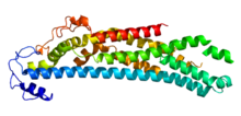
C-terminally truncated human glypican-1. PDB 4acr
|
|||||||||
| Identifiers | |||||||||
| Symbol | Glypican | ||||||||
| Pfam | PF01153 | ||||||||
| InterPro | IPR001863 | ||||||||
| PROSITE | PDOC00927 | ||||||||
|
|||||||||
| Available protein structures: | |
|---|---|
| Pfam | structures |
| PDB | RCSB PDB; PDBe; PDBj |
| PDBsum | structure summary |
Glypicans constitute one of the two major families of heparin sulfate proteoglycans, with the other major family being syndecans. Six glypicans have been identified in mammals, and are referred to as GPC1 through GPC6. In Drosophila two glypicans have been identified, and these are referred to as dally (division abnormally delayed) and dally-like. One additional glypican has been identified in C. elegans. Glypicans seem to play a vital role in developmental morphogenesis, and have been suggested as regulators for the Wnt and Hedgehog cell signaling pathways. They have additionally been suggested as regulators for fibroblast growth factor and bone morphogenic protein signaling.
While six glypicans have been identified in mammals, several characteristics remain consistent between these different proteins. First, the core protein of all glypicans is similar in size, approximately ranging between 60 and 70 kDa. Additionally, in terms of amino acid sequence, the location of fourteen cysteine residues is conserved; however, researchers describe glypicans as having moderate similarity in amino acid sequence overall. Nevertheless, it is thought that the fourteen conserved cysteine residues play a vital role in determining three-dimensional shape, thus suggesting the existence of a highly similar three-dimensional structure. Overall, GPC3 and GPC5 have very similar primary structures with 43% sequence similarity. On the other hand, GPC1, GPC2, GPC4, and GPC6 have between 35% and 63% sequence similarity. Thus, GPC3 and GPC5 are often referred to as one subfamily of glypicans, with GPC1, GPC2, GPC4, and GPC6 constituting the other group. Between the subfamilies of glypicans, there is about 25% sequence similarity. Furthermore, the amino acid sequence and structure of each glypican is well-conserved between species; it has been reported that all vertebrate glypicans are more than 90% similar regardless of the species.
For all members of the glypican family, the C-terminus of the protein is attached to the cell membrane covalently via a glycosylphosphatidylinositol (GPI) anchor. To allow for the addition of the GPI anchor, glypicans have a hydrophobic domain at the C-terminus of the protein. Within 50 amino acids of this GPI anchor, the heparan sulfate chains attach to the protein core. Therefore, unlike syndecans the heparan sulfate glycosaminoglycan chains attached to glypicans are located rather close to the cell-membrane. The glypicans found in vertebrates, Drosophila, and C. elegans all have an N-terminal signal sequence.
Glypicans are critically involved in developmental morphogenesis, and have been implicated as regulators in several cell signaling pathways. These include the Wnt and Hedgehog signaling pathways, as well as signaling of fibroblast growth factors and bone morphogenic proteins. The regulating processes performed by glypicans can either stimulate or inhibit specific cellular processes. The mechanisms by which glypicans regulate cellular pathways are not entirely elucidated. One commonly proposed mechanism suggests that glypicans behave as co-receptors which bind both the ligand and the receptor. Glypicans are expressed in various different amounts depending on the tissue, and they also are expressed to different degrees during the different stages of development. Drosophila studies have shown that dally mutants have irregular wing, antenna, genitalia, and brain development.
...
Wikipedia
