Gingival crevice
| Gingival sulcus | |
|---|---|

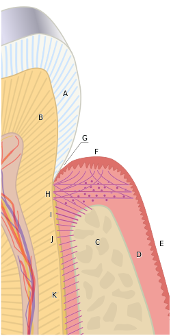
Gingival sulcus (G). Other letters: A, crown of the tooth, covered by enamel. B, root of the tooth, covered by cementum. C, alveolar bone. D, subepithelial connective tissue. E, oral epithelium. F, free gingival margin. H, principal gingival fibers. I, alveolar crest fibers of the PDL. J, horizontal fibers of the PDL. K, oblique fibers of the PDL.
|
|
| Details | |
| Identifiers | |
| Latin | sulcus gingivalis |
| TA | A05.1.01.111 |
| FMA | 74580 |
|
Anatomical terminology
[]
|
|
The gingival sulcus is an area of potential space between a tooth and the surrounding gingival tissue and is lined by sulcular epithelium. The depth of the sulcus (Latin for groove) is bounded by two entities: apically by the gingival fibers of the connective tissue attachment and coronally by the free gingival margin. A healthy sulcular depth is three millimeters or less, which is readily self-cleansable with a properly used toothbrush or the supplemental use of other oral hygiene aids.
When the sulcular depth is chronically in excess of three millimeters, regular home care may be insufficient to properly cleanse the full depth of the sulcus, allowing food debris and microbes to accumulate, forming dental biofilm. This poses a danger to the periodontal ligament (PDL) fibers that attach the gingiva to the tooth. If accumulated microbes remain undisturbed in a sulcus for an extended period of time, they will penetrate and ultimately destroy the delicate soft tissue and periodontal attachment fibers. If left untreated, this process may lead to a deepening of the sulcus, recession, destruction of the periodontium, including the bony tooth socket, tooth mobility, and tooth loss. A periodontal pocket is a dental term indicating the presence of an abnormally deepened gingival sulcus.
Rickne C. Scheid (2012). Woelfel's Dental Anatomy. Lippincott Williams & Wilkins. p. 200. ISBN .
...
Wikipedia
