Fuchs' dystrophy
| Fuchs' dystrophy | |
|---|---|
| Synonyms | Fuchs' corneal endothelial dystrophy (FCED) |
 |
|
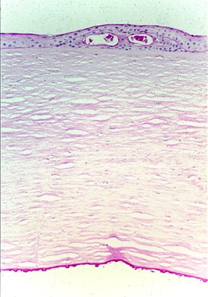
| Fuchs' corneal dystrophy. Light microscopic appearance of the cornea showing numerous excrescences (guttae) on the posterior surface of Descemet's membrane and the presence of cysts in the corneal epithelium beneath ectopically placed intraepithelial basement membrane. Periodic acid-Schiff stain. From a review by Klintworth, 2009. | |
| Pronunciation | |
| Classification and external resources | |
| Specialty | ophthalmology |
| ICD-10 | H18.5 |
| ICD-9-CM | 371.57 |
| OMIM | 136800 610158 |
| DiseasesDB | 31163 |
| MedlinePlus | 007295 |
| eMedicine | article/1193591 |
| MeSH | D005642 |
Fuchs' dystrophy is a slowly progressing corneal dystrophy that usually affects both eyes and is slightly more common in women than in men. Although doctors can often see early signs of Fuchs' dystrophy in people in their 30s and 40s, the disease rarely affects vision until people reach their 50s and 60s.
The condition was first described by Austrian ophthalmologist Ernst Fuchs (1851–1930), after whom it is named. In 1910, Fuchs first reported 13 cases of central corneal clouding, loss of corneal sensation and the formation of epithelial bullae, which he labeled ‘dystrophia epithelialis corneae’. It was characterized by late onset, slow progression, decreased visual acuity in the morning, lack of inflammation, diffuse corneal opacity, intense centrally, and roughened epithelium with vesicle-like features. A shift to the understanding of Fuchs’ corneal endothelial dystrophy (FCED) as primarily a disease of the corneal endothelium resulted after a number of observations in the 1920s. Crystal-like features of the endothelium were noted by Kraupa in 1920, who suggested that the epithelial changes were dependent on the endothelium. Using a slit lamp, Vogt described the excrescences associated with FCD as drop-like in appearance in 1921. In 1924, Graves then provided an extremely detailed explanation of the endothelial elevations visible with slit-lamp biomicroscopy. A patient with unilateral epithelial dystrophy and bilateral endothelial changes was described by the Friedenwalds in 1925; subsequent involvement of the second eye led them to emphasize that endothelial changes preceded epithelial changes. As only a subset of patients with endothelial changes proceeded to epithelial involvement, Graves stated on 19 October 1925 to the New York Academy of Medicine that “Fuchs’ epithelial dystrophy may be a very late sequel to severer cases of the deeper affection”.
FED may be discovered as an incidental finding at a routine optometrist visit or by an ophthalmologist during assessment for cataract surgery. As a result of irregularities on the inner surface of the cornea affected individuals may simply notice a reduction in the quality of vision or glare or haloes particularly when driving at night. Individuals with symptomatic Fuchs' dystrophy typically awaken with blurred vision which improves during the day. This occurs because the cornea is normally more swollen in the morning due to nocturnal fluid retention in the absence of normal evaporation due to the lids being closed. During waking hours this fluid evaporates once the eyes are open. As the disease worsens vision remains blurred despite evaporation due to endothelial pump failure and fluid retention. As Fuchs' dystrophy typically occurs in older individuals there may also be cataract of the lens which will also reduce vision.
...
Wikipedia
