Frontal nerve
| Frontal nerve | |
|---|---|

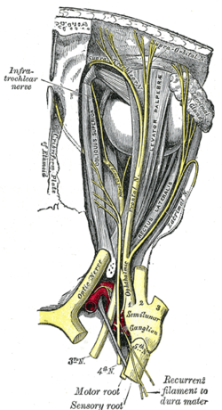
Dissection showing origins of right ocular muscles, and nerves entering by the superior orbital fissure.
|
|

Nerves of the orbit. Seen from above.
|
|
| Details | |
| From | Ophthalmic nerve |
| To | supratrochlear nerve and the supraorbital nerve |
| Identifiers | |
| Latin | nervus frontalis |
| TA | A14.2.01.020 |
| FMA | 52638 |
|
Anatomical terms of neuroanatomy
[]
|
|
The frontal nerve is the largest branch of the ophthalmic nerve(V1), and may be regarded, both from its size and direction, as the continuation of the nerve.
It enters the orbit through the superior orbital fissure, not running within the tendinous ring, and runs forward between the Levator palpebræ superioris and the periosteum.
Midway between the apex and base of the orbit it divides into two branches, supratrochlear nerve and supraorbital nerve.
It provides the sensory innervations for the skin of the forehead, mucosa of frontal sinus, and the skin of the upper eyelid via General Somatic Afferent (GSA) fibers.
Nerves of the orbit, and the ciliary ganglion. Side view.
Frontal nerve
Extrinsic eye muscle. Nerves of orbita. Deep dissection.
Extrinsic eye muscle. Nerves of orbita. Deep dissection.
Extrinsic eye muscle. Nerves of orbita. Deep dissection.
Extrinsic eye muscle. Nerves of orbita. Deep dissection.
Extrinsic eye muscle. Nerves of orbita. Deep dissection.
Extrinsic eye muscle. Nerves of orbita. Deep dissection.
Extrinsic eye muscle. Nerves of orbita. Deep dissection.
Frontal nerve.Deep dissection.Superior view
This article incorporates text in the public domain from the 20th edition of Gray's Anatomy (1918)
...
Wikipedia
