Foveæ
| Fovea centralis | |
|---|---|

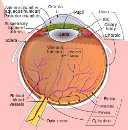
Schematic diagram of the human eye, with the fovea at the bottom. It shows a horizontal section through the right eye.
|
|
| Details | |
| Identifiers | |
| Latin | fovea centralis |
| TA | A15.2.04.022 |
| FMA | 58658 |
|
Anatomical terminology
[]
|
|
The fovea centralis (the term fovea comes from the Latin, meaning pit or pitfall) is a small, central pit composed of closely packed cones in the eye. It is located in the center of the macula lutea of the retina.
The fovea is responsible for sharp central vision (also called foveal vision), which is necessary in humans for activities where visual detail is of primary importance, such as reading and driving. The fovea is surrounded by the parafovea belt, and the perifovea outer region. The parafovea is the intermediate belt, where the ganglion cell layer is composed of more than five rows of cells, as well as the highest density of cones; the perifovea is the outermost region where the ganglion cell layer contains two to four rows of cells, and is where visual acuity is below the optimum. The perifovea contains an even more diminished density of cones, having 12 per 100 micrometres versus 50 per 100 micrometres in the most central fovea. This, in turn, is surrounded by a larger peripheral area that delivers highly compressed information of low resolution following the pattern of compression in foveated imaging.
Approximately half of the nerve fibers in the optic nerve carry information from the fovea, while the remaining half carry information from the rest of the retina. The parafovea extends to a radius of 1.25 mm from the central fovea, and the perifovea is found at a 2.75 mm radius from the fovea centralis.
Fovea (or Fovea centralis) is the depression in the inner retinal surface, about 1.5 mm wide, the photoreceptor layer of which is entirely cones and which is specialized for maximum visual acuity. Within the Fovea is a region of 0.5mm diameter celled the foveal avascular zone (which means without any blood vessels). This allows the light to be sensed without any dispersion or loss. This anatomy is responsible for the depression in the center of the fovea. The foveal pit is surrounded by the foveal rim that contains the neurons displaced from the pit. This is the thickest part of the retina.
...
Wikipedia
