Facial Nerve
| Facial nerve | |
|---|---|

Cranial nerve VII
|
|

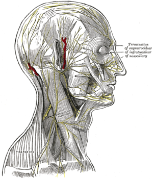
The nerves of the scalp, face, and side of neck.
|
|
| Details | |
| Identifiers | |
| Latin | nervus facialis |
| MeSH | A08.800.800.120.250 |
| Dorlands /Elsevier |
n_05/12565770 |
| TA | A14.2.01.099 |
| FMA | 50868 |
|
Anatomical terms of neuroanatomy
[]
|
|
The facial nerve is the seventh cranial nerve, or simply cranial nerve VII. It emerges from the brainstem between the pons and the medulla, controls the muscles of facial expression, and functions in the conveyance of taste sensations from the anterior two-thirds of the tongue and oral cavity. It also supplies preganglionic parasympathetic fibers to several head and neck ganglia.
The path of the facial nerve can be divided into six segments.
Distal to stylomastoid foramen, the following nerves branch off the facial nerve:
Intra operatively the facial nerve is recognized at 3 constant landmarks:
The cell bodies for the facial nerve are grouped in anatomical areas called nuclei or ganglia. The cell bodies for the afferent nerves are found in the geniculate ganglion for taste sensation. The cell bodies for muscular efferent nerves are found in the facial motor nucleus whereas the cell bodies for the parasympathetic efferent nerves are found in the superior salivatory nucleus.
The facial nerve is developmentally derived from the second pharyngeal arch, or branchial arch. The second arch is called the hyoid arch because it contributes to the formation of the lesser horn and upper body of the hyoid bone (the rest of the hyoid is formed by the third arch). The facial nerve supplies motor and sensory innervation to the muscles formed by the second pharyngeal arch, including the muscles of facial expression, the posterior belly of the digastric, stylohyoid and stapedius. The motor division of the facial nerve is derived from the basal plate of the embryonic pons, while the sensory division originates from the cranial neural crest.
...
Wikipedia
