Duct (anatomy)
| Duct | |
|---|---|

Dissection of a lactating breast.
1 - Fat 2 - Lactiferous duct/lobule 3 - Lobule 4 - Connective tissue 5 - Sinus of lactiferous duct 6 - Lactiferous duct |
|

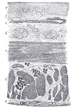
Section of the human esophagus. Moderately magnified. The section is transverse and from near the middle of the gullet.
a. Fibrous covering. b. Divided fibers of longitudinal muscular coat. c. Transverse muscular fibers. d. Submucous or areolar layer. e. Muscularis mucosae. f. Mucous membrane, with vessels and part of a lymphoid nodule. g. Stratified epithelial lining. h. Mucous gland. i. Gland duct. m’. Striated muscular fibers cut across. |
|
| Identifiers | |
| FMA | 30320 |
|
Anatomical terminology
[]
|
|
In anatomy and physiology, a duct is a circumscribed channel leading from an exocrine gland or organ.
Examples include:
As ducts travel from the acinus which generates the fluid to the target, the ducts become larger and the epithelium becomes thicker. The parts of the system are classified as follows:
Some sources consider "lobar" ducts to be the same as "interlobar ducts", while others consider lobar ducts to be larger and more distal from the acinus. For sources that make the distinction, the interlobar ducts are more likely to classified with simple columnar epithelium (or pseudostratified epithelium), reserving the stratified columnar for the lobar ducts.
Section of submaxillary gland of kitten. Duct semidiagrammatic. X 200.
Section of portion of mamma.
see also K.D.Tripathy...by K.E.R
...
Wikipedia
