Dermoscopy
| Dermatoscopy | |
|---|---|

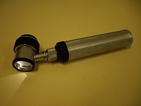
Immersion oil dermatoscope.
|
|
| MeSH | D046169 |
Dermatoscopy (also known as dermoscopy or epiluminescence microscopy) is the examination of skin lesions with a dermatoscope. This traditionally consists of a magnifier (typically x10), a non-polarised light source, a transparent plate and a liquid medium between the instrument and the skin, and allows inspection of skin lesions unobstructed by skin surface reflections. Modern dermatoscopes dispense with the use of liquid medium and instead use polarised light to cancel out skin surface reflections. When the images or video clips are digitally captured or processed, the instrument can be referred to as a "digital epiluminescence dermatoscope".
This instrument is useful to dermatologists in distinguishing benign from malignant (cancerous) lesions, especially in the diagnosis of melanoma.
With doctors who are experts in the specific field of dermoscopy, the diagnostic accuracy for melanoma is significantly better than for those dermatologists who do not have any specialized training in dermatoscopy. Thus, with specialists trained in dermoscopy, there is considerable improvement in the sensitivity (detection of melanomas) as well as specificity (percentage of non-melanomas correctly diagnosed as benign), compared with naked eye examination. The accuracy by dermatoscopy was increased up to 20% in the case of sensitivity and up to 10% in the case of specificity, compared with naked eye examination. By using dermatoscopy the specificity is thereby increased, reducing the frequency of unnecessary surgical excisions of benign lesions.
In the studies referred to, a comparison is made between dermatoscopy and an artificial situation of only naked eye examination. In reality, most of the 80% of US dermatologists who do not employ dermatoscopy work in bright rooms and use various magnifiers.
Skin surface microscopy started in 1663 by Kolhaus and was improved with the addition of immersion oil in 1878 by Ernst Abbe. The German dermatologist, Johann Saphier, added a built-in light source to the instrument. Goldman was the first dermatologist to coin the term "dermascopy" and to use the dermatoscope to evaluate pigmented cutaneous lesions.
In 2001, a California medical device manufacturer, 3Gen, introduced the first polarized dermatoscope, the DermLite. Polarised illumination, coupled with a cross-polarised viewer, reduces (polarised) skin surface reflection, thus allowing visualisation of skin structures (the light from which is depolarised) without using an immersion fluid. Examination of several lesions is thus more convenient because physicians no longer have to stop and apply immersion oil, alcohol, or water to the skin before examining each lesion. With the marketing of polarised dermatoscopes, dermatoscopy increased in popularity among physicians worldwide. Although images produced by polarised light dermatoscopes are slightly different from those produced by a traditional skin contact glass dermatoscope, they have certain advantages, such as vascular patterns not being potentially missed through compression of the skin by a glass contact plate.
...
Wikipedia
