Constrictor pharyngis superior
| Superior pharyngeal constrictor muscle | |
|---|---|

|
|

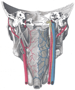
Muscles of the pharynx, viewed from behind, together with the associated vessels and nerves.
|
|
| Details | |
| Origin | Medial pterygoid plate, pterygomandibular raphé, alveolar process |
| Insertion | Pharyngeal raphe, pharyngeal tubercle |
| Artery | Ascending pharyngeal artery and tonsillar branch of facial artery |
| Nerve | Pharyngeal plexus of vagus nerve |
| Actions | Swallowing |
| Identifiers | |
| Latin | Musculus constrictor pharyngis superior |
| TA | A05.3.01.103 |
| FMA | 46621 |
|
Anatomical terms of muscle
[]
|
|
The superior pharyngeal constrictor muscle is a muscle in the pharynx. It is the highest located muscle of the three pharyngeal constrictors. The muscle is a quadrilateral muscle, thinner and paler than the inferior pharyngeal constrictor muscle and middle pharyngeal constrictor muscle.
The muscle is divided into four parts: A pterygopharyngeal, buccopharyngeal, mylopharyngeal and a glossopharyngeal part.
The four parts of this muscle arise from:
- the lower third of the posterior margin of the medial pterygoid plate and its hamulus (Pterygopharyngeal part)
- from the pterygomandibular raphe (Buccopharyngeal part)
- from the alveolar process of the mandible above the posterior end of the mylohyoid line (Mylopharyngeal part)
- and by a few fibers from the side of the tongue (Glossopharyngeal part)
The fibers curve backward to be inserted into the median raphe, being also prolonged by means of an aponeurosis to the pharyngeal spine on the basilar part of the occipital bone.
The superior fibers arch beneath the levator veli palatini muscle and the Eustachian tube.
The interval between the upper border of the muscle and the base of the skull is closed by the pharyngeal aponeurosis, and is known as the sinus of Morgagni.
...
Wikipedia
