Circumvallate papilla
| Lingual papillae | |
|---|---|

Anatomic landmarks of the tongue. Filiform papillae cover most of the dorsal surface of the anterior 2/3 of the tongue, with fungiform interspaced. Just in front of the sulcus terminalis lies a V-shaped line of circumvallate papillae, and on the posterior aspects of the lateral margins of the tongue lie the foliate papillae.
|
|

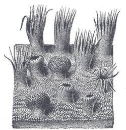
Semidiagrammatic view of a portion of the mucous membrane of the tongue. Two fungiform papillae are shown. On some of the filiform papillae the epithelial prolongations stand erect, in one they are spread out, and in three they are folded in.
|
|
| Details | |
| Identifiers | |
| Latin | papillae linguales GraySubject = 242 |
| Code | TH H3.04.01.0.03006 |
| Dorlands /Elsevier |
12610375 |
| TA | A05.1.04.013 |
| FMA | 54819 |
|
Anatomical terminology
[]
|
|
Lingual papillae (singular papilla) are the small, nipple-like structures on the upper surface of the tongue that give the tongue its characteristic rough texture. The four types of papillae on the human tongue have different structures and are named accordingly.
These are the: circumvallate papillae (vallate papillae), fungiform papillae, filiform papillae and foliate papillae. All except the filiform papillae are associated with taste buds.
In living subjects, lingual papillae are more readily seen when the tongue is dry. There are four types of papillae present on the tongue:
Filiform papillae are the most numerous of the lingual papillae. They are fine, small, cone-shaped papillae covering most of the dorsum of the tongue. They are responsible for giving the tongue its texture and are responsible for the sensation of touch. Unlike the other kinds of papillae, filiform papillae do not contain taste buds. They cover most of the front two-thirds of the tongue's surface.
They appear as very small, conical or cylindrical surface projections, and are arranged in rows which lie parallel to the sulcus terminalis. At the tip of the tongue, these rows become more transverse.
Histologically, they are made up of irregular connective tissue cores with a keratin–containing epithelium which has fine secondary processes. Heavy keratinization of filiform papillae, occurring for instance in cats, gives the tongue a roughness that is characteristic of these animals.
These processes have a whitish tint, owing to the thickness and density of their epithelium. This epithelium has undergone a peculiar modification as the cells have become cone–like and elongated into dense, overlapping, brush-like processes. They also contain a number of elastic fibers, which render them firmer and more elastic than the other types of papillae. The larger and longer papillae of this group are sometimes termed papillae conicae.
The fungiform papillae are mushroom shaped projections on the tongue,generally red in color. They are found on the upper surface of the tongue, scattered amongst the filiform papillae but are mostly present on the tip and sides of the tongue. They have taste buds on their upper surface which can distinguish the five tastes: sweet, sour, bitter, salty, and umami. They have a core of connective tissue. The fungiform papillae are innervated by the seventh cranial nerve, more specifically via the submandibular ganglion, chorda tympani, and geniculate ganglion ascending to the solitary nucleus in the brainstem.
...
Wikipedia
