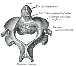
Cervical vertebra 2
| Axis | |
|---|---|

Position of axis (shown in red).
|
|

Second cervical vertebra, or epistropheus, from above.
|
|
| Details | |
| Identifiers | |
| Latin | Axis, vertebra cervicalis II |
| TA | A02.2.02.201 |
| FMA | 12520 |
|
Anatomical terms of bone
[]
|
|
In anatomy, the second cervical vertebra (C2) of the spine is named the axis (from Latin axis, "axle") or epistropheus.
By the atlanto-axial joint, it forms the pivot upon which the first cervical vertebra (the atlas), which carries the head, rotates.
The most distinctive characteristic of this bone is the strong odontoid process known as the dens which rises perpendicularly from the upper surface of the body. That peculiar feature gives to the vertebra a rarely used third name: vertebra dentata. In some judicial hangings the odontoid process may break and hit the medulla oblongata, causing death.
The body is deeper in front than behind, and prolonged downward anteriorly so as to overlap the upper and front part of the third vertebra.
It presents in front a median longitudinal ridge, separating two lateral depressions for the attachment of the Longus colli muscles.
Its under surface is concave from before backward and convex from side to side.
The dens, also odontoid process or peg, is the most pronounced feature, and exhibits a slight constriction or neck where it joins the main body of the vertebra. The dens is a protuberance (process or projection) of the axis (second cervical vertebra). The condition, where the dens is separated from the body of the axis, is called os odontoideum, and may cause nerve and circulation compression syndrome. On its anterior surface is an oval or nearly circular facet for articulation with that on the anterior arch of the atlas. On the back of the neck, and frequently extending on to its lateral surfaces, is a shallow groove for the transverse atlantal ligament which retains the process in position. The apex is pointed, and gives attachment to the apical odontoid ligament; below the apex the process is somewhat enlarged, and presents on either side a rough impression for the attachment of the alar ligament; these ligaments connect the process to the occipital bone.
...
Wikipedia
