Central auditory system
| Cochlea | |
|---|---|

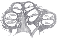
Diagrammatic longitudinal section of the cochlea. The cochlear duct, or scala media, is labeled as ductus cochlearis at right.
|
|
|
Anatomical terminology
[]
|
The auditory system is the sensory system for the sense of hearing. It includes both the sensory organs (the ears) and the auditory parts of the sensory system.
The outer ear funnels sound vibrations to the eardrum, increasing the sound pressure in the middle frequency range. The middle-ear ossicles further amplify the vibration pressure roughly 20 times. The base of the stapes couples vibrations into the cochlea via the oval window, which vibrates the perilymph liquid (present throughout the inner ear) and causes the round window to bulb out as the oval window bulges in. Vestibular and tympanic ducts are filled with perilymph, and the smaller cochlear duct between them is filled with endolymph, a fluid with a very different ion concentrations and voltage. Vestibular duct perilymph vibrations bend organ of Corti outer cells (4 lines) causing prestin to be released in cell tips. This causes the cells to be chemically elongated and shrunk (somatic motor), and hair bundles to shift which, in turn, electrically effects the basilar membrane’s movement (hair-bundle motor). These motors (outer cells) amplify the perilymph vibrations that initially incited them over 40-fold. Since both motors are chemically driven they are unaffected by the newly amplified vibrations due to recuperation time. The outer hair cells (OHC) are minimally innervated by spiral ganglion in slow (unmyelinated) reciprocal communicative bundles (30+ hairs per nerve fiber); this contrasts inner hair cells (IHC) that have only afferent innervation (30+ nerve fibers per one hair) but are heavily connected. There are 4x more OHC than IHC. The basilar membrane is a wall where the majority of the IHC and OHC sit. Basilar membrane width and stiffness corresponds to the frequencies best sensed by the IHC. At the cochlea base the Basilar is at its narrowest and most stiff (high-frequencies), at the cochlea apex it is at its widest and least stiff (low-frequencies). The tectorial membrane supports the remaining IHC and OHC. Tectorial membrane helps facilitate cochlear amplification by stimulating OHC (direct) and IHC (via endolymph vibrations). Tectorial's width and stiffness parallels Basilar's and similarly aids in frequency differentiation.
The superior olivary complex (SOC), in pons, is the first convergence of the left and right cochlear pulses. SOC has 14 described nuclei; their abbreviation are used here (see Superior olivary complex for their full names). MSO determines the angle the sound came from by measuring time differences in left and right info. LSO normalizes sound levels between the ears; it uses the sound intensities to help determine sound angle. LSO innervates the IHC. VNTB innervate OHC. MNTB inhibit LSO via glycine. LNTB are glycine-immune, used for fast signalling. DPO are high-frequency and tonotopical. DLPO are low-frequency and tonotopical. VLPO have the same function as DPO, but act in a different area. PVO, CPO, RPO, VMPO, ALPO and SPON (inhibited by glycine) are various signalling and inhibiting nuclei.
...
Wikipedia
