Carotid body
| Carotid body | |
|---|---|

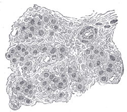
Section of part of human glomus caroticum. Highly magnified. Numerous blood vessels are seen in section among the gland cells.
|
|

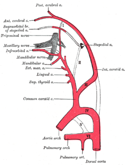
Diagram showing the origins of the main branches of the carotid arteries.
|
|
| Details | |
| Identifiers | |
| Latin | glomus caroticum |
| TA | A12.2.04.007 |
| FMA | 50095 |
|
Anatomical terminology
[]
|
|
The carotid body (carotid glomus or glomus caroticum) is a small cluster of chemoreceptors and supporting cells located near the fork (bifurcation) of the carotid artery (which runs along both sides of the throat).
The carotid body detects changes in the composition of arterial blood flowing through it, mainly the partial pressure of oxygen, but also of carbon dioxide. Furthermore, it is also sensitive to changes in pH and temperature.
The carotid body is made up of two types of cells, called glomus cells: glomus type I (chief) cells, and glomus type II (sustentacular cells).
The carotid body contains the most vascularized tissue in the human body. The thyroid gland is very vascular, but not quite as much as the carotid body.
The carotid body functions as a sensor: it responds to a stimulus, primarily O2 partial pressure, which is detected by the type I (glomus) cells, and triggers an action potential through the afferent fibers of the glossopharyngeal nerve, which relays the information to the central nervous system.
The carotid body chemoreceptors are primarily sensitive to decreases in the partial pressure of oxygen (PO2). This is in contrast to the central chemoreceptors in the medulla that are primarily sensitive to changes in pH and PCO2 (a decrease in pH and an increase in PCO2). The carotid body chemoreceptors are also sensitive to pH and PCO2, but only secondarily. More specifically, the sensitivity of carotid body chemoreceptors to decreased PO2 is greater when pH is decreased and PCO2 is increased.
The output of the carotid bodies is low at an oxygen partial pressure above about 100mmHg (13,3 kPa) (at normal physiological pH), but below 60mmHg the activity of the type I (glomus) cells increases rapidly due to a decrease in hemoglobin-oxygen saturation below 90%.
...
Wikipedia
