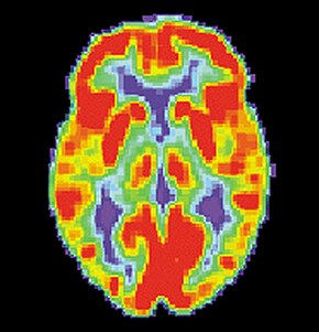
Brain positron emission tomography
| Brain positron emission tomography | |
|---|---|
| Medical diagnostics | |

PET scan of a normal brain
|
|
| ICD-10-PCS | C030 |
Positron emission tomography (PET) measures emissions from radioactively labeled metabolically active chemicals that have been injected into the bloodstream. The emission data are computer-processed to produce multi-dimensional images of the distribution of the chemicals throughout the brain.
The positron emitting radioisotopes used are usually produced by a cyclotron, and chemicals are labeled with these radioactive atoms. The labeled compound, called a radiotracer, is injected into the bloodstream and eventually makes its way to the brain through blood circulation. Detectors in the PET scanner detect the radioactivity as the compound charges in various regions of the brain. A computer uses the data gathered by the detectors to create multi-dimensional (normally 3-dimensional volumetric or 4-dimensional time-varying) images that show the distribution of the radiotracer in the brain. Especially useful are a wide array of ligands used to map different aspects of neurotransmitter activity, with by far the most commonly used PET tracer being a labeled form of glucose (see Fludeoxyglucose (18F) (FDG)).
The greatest benefit of PET scanning is that different compounds can show blood flow and oxygen and glucose metabolism in the tissues of the working brain. These measurements reflect the amount of brain activity in the various regions of the brain and allow to learn more about how the brain works. PET scans were superior to all other metabolic imaging methods in terms of resolution and speed of completion (as little as 30 seconds), when they first became available. The improved resolution permitted better study to be made as to the area of the brain activated by a particular task. The biggest drawback of PET scanning is that because the radioactivity decays rapidly, it is limited to monitoring short tasks.
Before the use of Functional magnetic resonance imaging became widespread, PET scanning was the preferred method of functional (as opposed to structural) brain imaging, and it still continues to make large contributions to neuroscience. PET scanning is also useful in PET-guided stereotactic surgery and radiosurgery for treatment of intracranial tumors, arteriovenous malformations and other surgically treatable conditions.
...
Wikipedia
