Black lung disease
| Coalworker's pneumoconiosis | |
|---|---|
 |
|
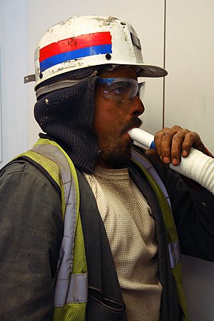
| A coal miner performing spirometry to assess his lung function | |
| Classification and external resources | |
| Specialty | pulmonology |
| ICD-10 | J60 |
| ICD-9-CM | 500 |
| DiseasesDB | 10145 |
| MedlinePlus | 000130 |
| eMedicine | med/398 |
| MeSH | D011009 |
Coal workers' pneumoconiosis (CWP), also known as black lung disease or black lung, is caused by long exposure to coal dust. It is common in coal miners and others who work with coal. It is similar to both silicosis from inhaling silica dust and to the long-term effects of tobacco smoking. Inhaled coal dust progressively builds up in the lungs and cannot be removed by the body; this leads to inflammation, fibrosis, and in worse cases, necrosis.
Coal workers' pneumoconiosis, severe state, develops after the initial, milder form of the disease known as anthracosis (anthrac — coal, carbon). This is often asymptomatic and is found to at least some extent in all urban dwellers due to air pollution. Prolonged exposure to large amounts of coal dust can result in more serious forms of the disease, simple coal workers' pneumoconiosis and complicated coal workers' pneumoconiosis (or progressive massive fibrosis, or PMF). More commonly, workers exposed to coal dust develop industrial bronchitis, clinically defined as chronic bronchitis (i.e. productive cough for 3 months per year for at least 2 years) associated with workplace dust exposure. The incidence of industrial bronchitis varies with age, job, exposure, and smoking. In nonsmokers (who are less prone to develop bronchitis than smokers), studies of coal miners have shown a 16% to 17% incidence of industrial bronchitis.
In 2013 CWP resulted in 25,000 deaths down from 29,000 deaths in 1990.
Coal dust is not as fibrogenic as in silica dust. Coal dust that enters the lungs can neither be destroyed nor removed by the body. The particles are engulfed by resident alveolar or interstitial macrophages and remain in the lungs, residing in the connective tissue or pulmonary lymph nodes. Coal dust provides a sufficient stimulus for the macrophage to release various products, including enzymes, cytokines, oxygen radicals, and fibroblast growth factors, which are important in the inflammation and fibrosis of CWP. Aggregations of carbon-laden macrophages can be visualized under a microscope as granular, black areas. In serious cases, the lung may grossly appear black. These aggregations can cause inflammation and fibrosis, as well as the formation of nodular lesions within the lungs. The centers of dense lesions may become necrotic due to ischemia, leading to large cavities within the lung.
...
Wikipedia
