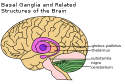
Basal nuclei
| Basal ganglia | |
|---|---|

Basal ganglia labeled at top right.
|
|

Basal ganglia on underneath view of brain
|
|
| Details | |
| Part of | Cerebrum |
| Identifiers | |
| Latin | nuclei basales |
| MeSH | A08.186.211.730.885.105 |
| NeuroNames | hier-206 |
| NeuroLex ID | Basal ganglia |
| Dorlands /Elsevier |
n_11/12580456 |
| TA | A14.1.09.501 |
| FMA | 84013 |
|
Anatomical terms of neuroanatomy
[]
|
|
The basal ganglia (or basal nuclei) is a group of subcortical nuclei, of varied origin, in the brains of vertebrates including humans, which are situated at the base of the forebrain. Basal ganglia nuclei are strongly interconnected with the cerebral cortex, thalamus, and brainstem, as well as several other brain areas. The basal ganglia are associated with a variety of functions including: control of voluntary motor movements, procedural learning, routine behaviors or "habits" such as teeth grinding, eye movements, cognition, and emotion.
The main components of the basal ganglia – as defined functionally – are the striatum both dorsal striatum (caudate nucleus and putamen) and ventral striatum (nucleus accumbens and olfactory tubercle), globus pallidus, ventral pallidum, substantia nigra, and subthalamic nucleus. Each of these components has a complex internal anatomical and neurochemical organization. The largest component, the striatum (dorsal and ventral), receives input from many brain areas beyond the basal ganglia, but only sends output to other components of the basal ganglia. The pallidum receives input from the striatum, and sends inhibitory output to a number of motor-related areas. The substantia nigra is the source of the striatal input of the neurotransmitter dopamine, which plays an important role in basal ganglia function. The subthalamic nucleus receives input mainly from the striatum and cerebral cortex, and projects to the globus pallidus.
...
Wikipedia
