Area postrema
| Area postrema | |
|---|---|

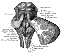
Rhomboid fossa. (Area postrema labeled at bottom center.)
|
|

Human caudal brainstem posterior view description (Area postrema is #8)
|
|
| Identifiers | |
| Acronym(s) | AP |
| MeSH | A08.186.211.132.810.406.286 |
| NeuroNames | hier-769 |
| NeuroLex ID | Area Postrema |
| TA | A14.1.04.258 |
| FMA | 72607 |
|
Anatomical terms of neuroanatomy
[]
|
|
The area postrema is a medullary structure in the brain that controls vomiting. Its privileged location in the brain also allows the area postrema to play a vital role in the control of autonomic functions by the central nervous system.
The area postrema is a small protuberance found at the inferoposterior limit of the fourth ventricle. Specialized ependymal cells are found within the area postrema. These specialized ependymal cells differ slightly from the majority of ependymal cells (ependymocytes), forming a unicellular epithelium lining of the ventricles and central canal. The area postrema is separated from the vagal triangle by the funiculus separans, a thin semitransparent ridge. The vagal triangle overlies the dorsal vagal nucleus and is situated on the caudal end of the rhomboid fossa or 'floor' of the fourth ventricle. The area postrema is situated just before the obex, the inferior apex of the caudal ventricular floor. Both the funiculus separans and area postrema have a similar thick ependyma-containing tanycyte covering. Ependyma and tanycytes can participate in transport of neurochemicals into and out of the cerebrospinal fluid from its cells or adjacent neurons, glia or vessels. Ependyma and tanycytes may also participate in chemoreception. The eminence of the area postrema is considered a circumventricular organ because its endothelial cells do not contain tight junctions, which allows for free exchange of molecules between blood and brain tissue. This unique breakdown in the blood–brain barrier is partially compensated for by the presence of a tanycyte barrier.
The area postrema connects to the nucleus of the solitary tract and other autonomic control centers in the brainstem. It is excited by visceral afferent impulses (sympathetic and vagal) arising from the gastrointestinal tract and other peripheral trigger areas. The area postrema makes up part of the dorsal vagal complex, which is the critical termination site of vagal afferent nerve fibers, along with the dorsal motor nucleus of the vagus and the nucleus of the solitary tract. Vomiting and nausea are most likely induced by the area postrema through its connection to the nucleus of the solitary tract, which may serve as the beginning of the pathway triggering vomiting in response to various emetic inputs. However, this structure plays no key role for vomiting induced by the activation of vagal nerve fibers or by motion, and its function in radiation-induced vomiting remains unclear. Because the area postrema is located outside of the blood–brain barrier, peptide and other physiological signals in the blood have direct access to neurons of brain areas with vital roles in the autonomic control of the body. As a result, the area postrema is now being considered as the initial site for integration for various physiological signals in the blood as they enter the central nervous system.
...
Wikipedia
