Anterior arch of atlas
| Atlas (anatomy) | |
|---|---|

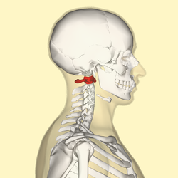
Position of the atlas (shown in red)
|
|
| Details | |
| Identifiers | |
| Latin | Atlas, vertebra cervicalis I |
| Dorlands /Elsevier |
a_70/12167134 |
| TA | A02.2.02.101 |
| FMA | 12519 |
|
Anatomical terms of bone
[]
|
|
In anatomy, the atlas (C1) is the most superior (first) cervical vertebra of the spine.
It is named for the Atlas of Greek mythology, because it supports the globe of the head.
The atlas is the topmost vertebra and with the axis forms the joint connecting the skull and spine. The atlas and axis are specialized to allow a greater range of motion than normal vertebrae. They are responsible for the nodding and rotation movements of the head.
The atlanto-occipital joint allows the head to nod up and down on the vertebral column. The dens acts as a pivot that allows the atlas and attached head to rotate on the axis, side to side.
The atlas's chief peculiarity is that it has no body. It is ring-like and consists of an anterior and a posterior arch and two lateral masses.
The atlas and axis are important neurologically because the brain stem extends down to the axis.
The anterior arch forms about one-fifth of the ring: its anterior surface is convex, and presents at its center the anterior tubercle for the attachment of the Longus colli muscles and the anterior longitudinal ligament; posteriorly it is concave, and marked by a smooth, oval or circular facet (fovea dentis), for articulation with the odontoid process (dens) of the axis.
The upper and lower borders respectively give attachment to the anterior atlantooccipital membrane and the anterior atlantoaxial ligament; the former connects it with the occipital bone above, and the latter with the axis below.
...
Wikipedia
