Angular cheilitis
| Angular cheilitis | |
|---|---|
| Synonyms | rhagades, perlèche, cheilosis, angular cheilosis, commissural cheilitis, angular stomatitis |
 |
|
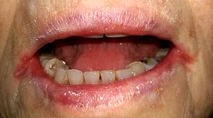
| Bilateral angular cheilitis in an elderly individual with false teeth, iron deficiency anemia and xerostomia | |
| Pronunciation | /kaɪˈlaɪtɪs/ |
| Classification and external resources | |
| Specialty | Dermatology |
| ICD-10 | K13.0 |
| ICD-9-CM | 528.5, 686.8 |
| MeSH | D002613 |
Angular cheilitis (AC), is inflammation of one or both corners of the mouth. Often the corners are red with skin breakdown and crusting. It can also be itchy or painful. The condition can last for days to years. Angular cheilitis is a type of cheilitis (inflammation of the lips).
Angular cheilitis can be caused by infection, irritation, or allergies. Infections include by the fungi such as Candida albicans and bacteria such as Staph. aureus. Irritants include poorly fitting dentures, licking the lips or drooling, mouth breathing resulting in a dry mouth, sun exposure, overclosure of the mouth, smoking, and minor trauma. Allergies may include to substances like toothpaste, makeup, and food. Often a number of factors are involved. Other factors may include poor nutrition or poor immune function. Diagnosis may be helped by testing for infections and patch testing for allergies.
Treatment for angular cheilitis is typically based on the underlying causes along with the use of a barrier cream. Frequently an antifungal and antibacterial cream is also tried. Angular cheilitis is a fairly common problem, with estimates that it affects 0.7% of the population. It occurs most often in the 30s to 60s, although is also relatively common in children. In the developing world, iron and vitamin deficiencies are a common cause.
Angular cheilitis is a fairly non specific term which describes the presence of an inflammatory lesion in a particular anatomic site (i.e. the corner of the mouth). As there are different possible causes and contributing factors from one person to the next, the appearance of the lesion is somewhat variable. The lesions are more commonly symmetrically present on both sides of the mouth, but sometimes only one side may be affected. In some cases, the lesion may be confined to the mucosa of the lips, and in other cases the lesion may extend past the vermilion border (the edge where the lining on the lips becomes the skin on the face) onto the facial skin. Initially, the corners of the mouth develop a gray-white thickening and adjacent erythema (redness). Later, the usual appearance is a roughly triangular area of erythema, edema (swelling) and breakdown of skin at either corner of the mouth. The mucosa of the lip may become fissured (cracked), crusted, ulcerated or atrophied. There is not usually any bleeding. Where the skin is involved, there may be radiating rhagades (linear fissures) from the corner of the mouth. Infrequently, the dermatitis (which may resemble eczema) can extend from the corner of the mouth to the skin of the cheek or chin. If Staphylococcus aureus is involved, the lesion may show golden yellow crusts. In chronic angular cheilitis, there may be suppuration (pus formation), exfoliation (scaling) and formation of granulation tissue.
...
Wikipedia
