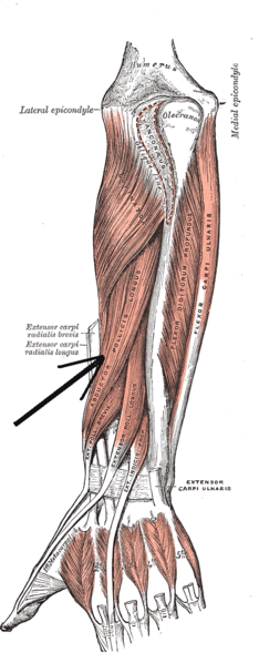
Abductor pollicis longus
| Abductor pollicis longus muscle | |
|---|---|

Deep muscles of posterior surface of the forearm
|
|
| Details | |
| Origin | ulna, radius, Interosseous membrane |
| Insertion | 1st metacarpal |
| Artery | Posterior interosseous artery |
| Nerve |
Posterior interosseous nerve from deep branch of radial nerve C7, C8 |
| Actions | abduction, extension of thumb |
| Antagonist | Adductor pollicis muscle |
| Identifiers | |
| Latin | musculus abductor pollicis longus |
| TA | A04.6.02.049 |
| FMA | 38515 |
|
Anatomical terms of muscle
[]
|
|
In human anatomy, the abductor pollicis longus (APL) is one of the extrinsic muscles of the hand. As the name implies, its major function is to abduct the thumb at the wrist. Its tendon forms the anterior border of the anatomical snuffbox.
The abductor pollicis longus lies immediately below the supinator and is sometimes united with it. It arises from the lateral part of the dorsal surface of the body of the ulna below the insertion of the anconeus, from the interosseous membrane, and from the middle third of the dorsal surface of the body of the radius.
Passing obliquely downward and lateralward, it ends in a tendon, which runs through a groove on the lateral side of the lower end of the radius, accompanied by the tendon of the extensor pollicis brevis.
The insertion is divided into a distal, superficial part and a proximal, deep part. The superficial part is inserted with one or more tendons into the radial side of the base of the first metacarpal bone, and the deep part is variably inserted into the trapezium, the joint capsule and its ligaments, and into the belly of abductor pollicis brevis (APB) or opponens pollicis.
The abductor pollicis longus muscle is innervated by the posterior interosseous nerve, which is a continuation of the deep branch of the radial nerve after it passes through the supinator muscle. The posterior interosseous nerve is derived from spinal segments C7 & C8.
Abductor pollicis longus is supplied by the posterior interosseous artery.
An accessory abductor pollicis longus (AAPL) tendon is present in more than 80% of people and a separate muscle belly is present in 20% of people. In one study, the accessory tendon was inserted into the trapezium (41%); proximally on the abductor pollicis brevis (22%) and opponens pollicis brevis (5%); had a double insertion on the trapezium and thenar muscles (15%); or the base of the first metacarpal (1%). Up to seven tendons have been reported in rare cases.
...
Wikipedia
