Zygomatic bone
| Zygomatic bone | |
|---|---|

Left zygomatic bone in situ.
|
|

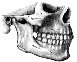
Side view of the teeth and jaws. (Zygomatic visible in center.)
|
|
| Details | |
| Identifiers | |
| Latin | os zygomaticum, zygoma |
| TA | A02.1.14.001 |
| FMA | 52747 |
|
Anatomical terms of bone
[]
|
|
In the human skull, the zygomatic bone (cheekbone or malar bone) is a paired bone which articulates with the maxilla, the temporal bone, the sphenoid bone and the frontal bone. It is situated at the upper and lateral part of the face and forms the prominence of the cheek, part of the lateral wall and floor of the orbit, and parts of the temporal and infratemporal fossa. It presents a malar and a temporal surface; four processes, the frontosphenoidal, orbital, maxillary, and temporal; and four borders.
The malar surface is convex and perforated near its center by a small aperture, the zygomaticofacial foramen, for the passage of the zygomaticofacial nerve and vessels; below this foramen is a slight elevation, which gives origin to the zygomaticus muscle.
The temporal surface, directed posteriorly and medially, is concave, presenting medially a rough, triangular area, for articulation with the maxilla (articular surface), and laterally a smooth, concave surface, the upper part of which forms the anterior boundary of the temporal fossa, the lower a part of the infratemporal fossa. Near the center of this surface is the zygomaticotemporal foramen for the transmission of the zygomaticotemporal nerve.
The orbital surface forms the lateral part and some of the inferior part of the bony orbit. The zygomatic nerve passes through the zygomatic-orbital foramen on this surface. The lateral palpebral ligament attaches to a small protuberance called the orbital tubercle.
Each zygomatic bone is diamond-shaped and composed of three processes with similarly named associated bony articulations: frontal, temporal, and maxillary. Each process of the zygomatic bone forms important structures of the skull.
The orbital surface of the frontal process of the zygomatic bone forms the anterior lateral orbital wall, with usually a small paired foramen, the zygomaticofacial foramen opening on its lateral surface. The temporal process of the zygomatic bone forms the zygomatic arch along with the zygomatic process of the temporal bone, with a paired zygomaticotemporal foramen present on the medial deep surface of the bone. The orbital surface of the maxillary process of the zygomatic bone forms a part of the infraorbital rim and a small part of the anterior part of the lateral orbital wall.
...
Wikipedia
