Vein
| Vein | |
|---|---|

The main veins in the human body
|
|

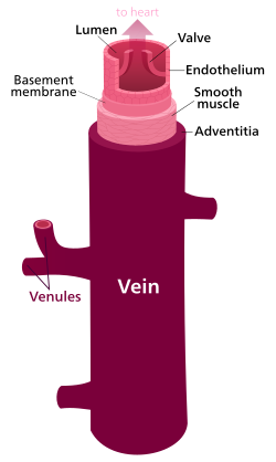
Structure of a vein, which consists of three main layers. The outer layer is connective tissue, called tunica adventitia or tunica externa; a middle layer of smooth muscle called the tunica media, and the inner layer lined with endothelial cells called the tunica intima.
|
|
| Details | |
| System | Circulatory system |
| Identifiers | |
| Latin | vena |
| TA |
A12.0.00.030 A12.3.00.001 |
| FMA | 50723 |
|
Anatomical terminology
[]
|
|
Veins are blood vessels that carry blood toward the heart. Most veins carry deoxygenated blood from the tissues back to the heart; exceptions are the pulmonary and umbilical veins, both of which carry oxygenated blood to the heart. In contrast to veins, arteries carry blood away from the heart.
Veins are less muscular than arteries and are often closer to the skin. There are valves in most veins to prevent backflow.
Veins are present throughout the body as tubes that carry blood back to the heart. Veins are classified in a number of ways, including superficial vs. deep, pulmonary vs. systemic, and large vs. small.
Most veins are equipped with valves to prevent blood flowing in the reverse direction.
Veins are translucent, so the color a vein appears from an organism's exterior is determined in large part by the color of venous blood, which is usually dark red as a result of its low oxygen content. Veins appear blue because the subcutaneous fat absorbs low-frequency light, permitting only the highly energetic blue wavelengths to penetrate through to the dark vein and reflect back to the viewer. The colour of a vein can be affected by the characteristics of a person's skin, how much oxygen is being carried in the blood, and how big and deep the vessels are. When a vein is drained of blood and removed from an organism, it appears grey-white.
The largest veins in the human body are the venae cavae. These are two large veins which enter the right atrium of the heart from above and below. The superior vena cava carries blood from the arms and head to the right atrium of the heart, while the inferior vena cava carries blood from the legs and abdomen to the heart. The inferior vena cava is retroperitoneal and runs to the right and roughly parallel to the abdominal aorta along the spine. Large veins feed into these two veins, and smaller veins into these. Together this forms the venous system.
...
Wikipedia
