Paraventricular nucleus
| Paraventricular nucleus of hypothalamus | |
|---|---|

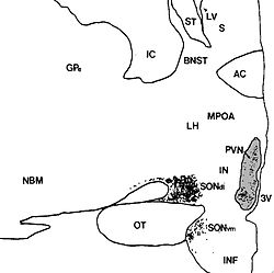
Human paraventricular nucleus (PVN) in this coronal section is indicated by the shaded area. Dots represent vasopressin (AVP) neurons (also seen in the supraoptic nucleus, SON). The medial surface is the 3rd ventricle (3V).
|
|

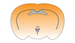
The paraventricular hypothalamus of the mouse brain
|
|
| Details | |
| Identifiers | |
| Latin | Nucleus paraventricularis hypothalami |
| MeSH | A08.186.211.730.385.357.342.400 |
| NeuroNames | hier-370 |
| NeuroLex ID | Paraventricular nucleus of hypothalamus |
| Dorlands /Elsevier |
n_11/12582324 |
| TA | A14.1.08.909 |
| FMA | 62320 |
|
Anatomical terms of neuroanatomy
[]
|
|
The paraventricular nucleus (PVN, PVA, or PVH) is a neuronal nucleus in the hypothalamus. It contains groups of neurons that can be activated by stressful and/or physiological changes. Many PVN neurons project directly to the posterior pituitary where they release into the general circulation.While the Supraoptic nucleus release vasopressin. Other PVN neurons control various anterior pituitary functions, while still others directly regulate appetite and autonomic functions in the brainstem and spinal cord.
The paraventricular nucleus lies adjacent to the third ventricle. It lies within the periventricular zone and is not to be confused with the periventricular nucleus, which occupies a more medial position, beneath the third ventricle. The PVN is highly vascularised and is protected by the blood–brain barrier, although its neuroendocrine neurons extend to sites (in the median eminence and in the posterior pituitary) beyond the blood–brain barrier.
The PVN contains magnocellular neurosecretory cells whose axons extend into the posterior pituitary, parvocellular neurosecretory (neuroendocrine) cells that project to the median eminence, and several populations of peptide-containing cells that project to many different brain regions including parvocellular preautonomic cells that project to the brainstem and spinal cord.
...
Wikipedia
